


1. Introduction
The electric signals associated with cells facilitate various biological processes in living bodies. Nerve cells are able to concentrate ions and generate a membrane potential, with a high difference of charge between the inside and outside of the cell, and rapid de-polarization enables a rapid transmission of information along a nerve fiber [1]. It has been reported that electrical energy emanating from sources outside the body can also affect cellular responses, such as cell migration and proliferation [2,3]. It has been observed that there is an electrical gradient between tumor and normal tissues and that malignant and normal cells have differential sensitivities for electric energy [4]. Based upon these results, a new line of research is developing that aims to use electric fields or currents in the localized treatment of malignant tumors [5-7].
We have shown that electric polarization treatment on bioceramics, such as hydroxyapatite and titania, produced electric fields around the material and induced surface charges without even microscopic changes of the surface morphology or alterations of the chemical composition [8,9]. Because these polarized ceramic biomaterials can be charged with electric energy before implantation inside the body, large-scale equipment is not needed at the point of use. The electrostatic energy stored in the polarized apatite and/or titania has been reported to control protein adsorption, to modify initial osteoblast responses, and to promote bone healing, including osteoblast proliferation and differentiation into osteocytes [10-13]. Also seen in animal models, polarization inhibited neointimal hyperplasia after angioplasty, by reducing the proliferation of endothelial cells and influencing the differentiation of smooth muscle cells [14]. These results indicate that the electric signals produced by surface fields around bioceramics can affect the balance of cell proliferation and differentiation. The coordination of these two cellular processes is especially important for the growth and maintenance of stem cells that are able to divide while maintaining their pluripotency [15]. To control the balance in malignant tumors could lead to suppress their uncontrolled expansion.
The current treatment for leiomyosarcoma is usually a combination of tumorectomy, chemotherapy, and radiotherapy. Despite improvements in these treatments, the prognosis of this solid tumor disease is still poor due to high rates of local recurrence and distant metastasis. An adjunct therapy that has an orthogonal anti-tumor action and can be used in conjunction with those therapies must be developed to improve the outcome for this disease. Recently, the new approach of differentiation-inducing therapy, in which tumor cells are influenced to differentiate into mature cells has been applied in a clinical setting. Specifically, all-trans retinoic acid was used to treat acute promyelocytic leukemia [16-18]. The malignancy of leiomyosarcoma may be reduced by using extrinsic influences, such as electric fields to switch the smooth muscle cells to a differentiated phenotype with a slower growth rate [19].
The molecular basis of the switch between replication as a stem cell and adoption of a particular cellular fate has been recently analyzed using cell cycle research in a cross-disciplinary manner [20,21]. In the present study, we examined the effects of the electric energy of a polarized apatite growth surface on the regulation of proliferation and differentiation of tumor cells, using the human uterine leiomyosarcoma cell line SK-LMS-1 (SK). This cell line has become immortalized, making it similar to stem or tumor cells, but can still be differentiated by some external signals, and its status can be evaluated by markers of differentiation, such as calponin and caldesmon [22,23]. Furthermore, the molecular mechanism of cell fate regulation, as affected by growth on a polarized surface, was investigated by examining changes in the expression of cell cycle-related genes.
2. Materials and Method
2.1 Preparation of specimens
Hydroxylapatite (HAp) powders synthesized by a wet method were uniaxially pressed into pellets. Then they were sintered at 1250°C for 2 h under a water vapor stream [24]. The specimen, a disk 10 mm in diameter and 0.8 mm thick, was sandwiched between a pair of platinum (Pt) plate electrodes, electrically polarized in a DC field of 5 kV·cm-1 in an air atmosphere at 400°C for 1 h, and then cooled to room temperature under the applied DC voltage [24,25]. The surface of specimens in contact with the cathode was labeled the negative surface (N-surface), and the surface placed in contact with the anode was labeled the positive surface (P-surface). A non-polarized specimen was heated in air at 400°C for 1 h with no electric field and labeled the 0-surface.
2.2 Characterization of the specimens
The crystal phase of the specimen was determined with a powder X-ray diffractometer (XRD, Bruker AXS D8 Advance) using Cu Kα radiation. Fourier transform infrared spectroscopy (FT-IR) was used to analyze the HAp specimens using the KBr pellet method (JASCO FT/IR-500 spectrometer). The thermally stimulated depolarization current (TSDC) of the polarized samples was measured using a picoampere meter (Keithley Inc., OH, USA). Each polarized sample was sandwiched between Pt electrodes and heated to 670°C in an electric furnace equipped with a stainless-steel shielded sample chamber at a heating rate of 5°C min-1. The stored charge was calculated by integration of the current density on the measurement as previously reported [26].
2.3 Cell culture
The human vulvar uterine leiomyosarcoma cell line, SK-LMS-1 (American Type Culture Collection) was grown in DMEM supplemented with 10% fetal bovine serum, 1% non-essential amino acids, and 1% Penicillin/Streptomycin.
2.4 Cell proliferation assay and apoptosis assay
Cell viability was quantitatively analyzed by using an MTT assay, which is widely used for measuring cytotoxity and proliferation. After seeding for 1, 3, or 6 days, the specimens were incubated with an MTT solution (5 mg/ml, Dojindo, Japan) at 37°C for 3 h. The formazan product, which was obtained by the reduction of MTT in the mitochondria of viable cells, was dissolved in dimethyl sulfoxide for 10 min and the absorbance was measured at 570 nm using a microplate reader (Bio-Rad, CA, USA). The DeadEnd™ Colorimetric Apoptosis Detection System (Promega, IL, USA) was used to visualize fragmented DNA arising from apoptotic cells. The terminal deoxynucleotidyl transferase deoxy-UTP nick end labeling (TUNEL)-positive cells in the resulting digital images (Olympus IX71 fluorescence microscope and DP71 digital camera; Olympus, Tokyo, Japan) were counted using MetaMorph software (Molecular Devices, CA, USA).
2.5 Measurement of gene expression by quantitative real-time reverse transcription-polymerase chain reaction (qRT-PCR) analysis
Total RNA from cells cultured for 24 h was extracted using spin columns according to the manufacturer’s instructions (Quick-RNA Micro Prep Kit, Zymo Research, Irvine, CA). Two micrograms of total RNA was used in transcription to create cDNA using SuperScript VILO according to the manufacturer’s protocol (Invitrogen). Then, the synthesized cDNA was used directly in qRTPCR analyses with the LightCycler System (Roche Applied Science, IN, USA). Amplifications were performed using a hydrolysis probe and primers obtained from the Human Universal ProbeLibrary (Roche) for selective genes (N = 4, the primers and universal probes are listed in Table 1) or RealTime ready Human Cell Cycle Regulation Panel for the array analysis in combination with LightCycler 480 Probes Master (Roche). Results were quantified based on the threshold cycle (Ct) value according to the 2-ΔΔCt method and normalized using the reference gene GAPDH. The fold changes in expression were compared between samples from polarized and non-polarized specimens. Increases of more than 1.75- fold and decreases to less than 0.75-fold were defined as substantial.
2.6 Statistical analysis
All the values are presented as the means ± standard deviation. Statistical analyses were performed using one-way analysis of variance followed by a post-hoc test for multiple comparisons (Ryan's test) with P < 0.05 considered statistically significant (ANOVA4 on the Web, Japan).
3. Results
3.1 Characterization of the polarized HAp
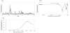
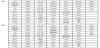
All specimens have more than 95% of the theoretical density value of HAp (3.16 g/cm3). The results of XRD and FT-IR analyses indicated that all the specimens consisted of a single apatitic phase of crystalline HAp (JCPDS 09-0432) and the IR absorption spectra of the specimens showed the typical absorption bands corresponding to the modes of phosphate at 1074 cm-1/ 1044 cm-1(ν3), 962 cm-1 (ν1), and 601 cm-1/570 cm-1 (ν4) and hydroxyl at 3569 cm-1 and 630 cm-1 (Figure 1a and Figure 1b). In addition, there were no obvious changes in morphology or phase composition after electric polarization. The average stored charges of the polarized specimens calculated from the TSDC curves were 2.8 μC.cm-2 , and the charges of non-polarized specimens were too low to be measured (Figure 1c).
3.2 Cell proliferation, apoptosis, and differentiation
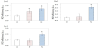
SK cells grown on culture glass, on polarized apatite, or on control non-polarized apatite, were evaluated by MTT assays 1, 3, or 6 d after seeding (Figure 2a and Figure 2b). The proliferation rates of the cells on all substrata increased with time. Although there were no significant differences among the three surfaces at 1 d and 3 d, the proliferation rates of cells on the polarized specimens decreased at 6 d compared to the non-polarized surface.
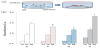
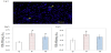
To examine the reasons for the differences of the proliferation rates among the specimens, an apoptosis assay was performed that detected the number of TUNEL-positive cells (Figure 3a). There were a small number of apoptotic cells on the apatite specimens with or without polarization. Then the degree of differentiation was evaluated using real-time reverse transcriptase PCR. Calponin and caldesmon, markers specific to smooth muscle cells, can reflect the degree of differentiation of SK cells. The expression levels of these two genes were ranked highest to lowest for cells grown on the P-surface, N-surface, and 0-surface (Figure 3b and Figure 3c). The results seemed to correlate with a ranking according to decreasing proliferation rates(greater decreases on the polarized surfaces than on the 0-surface).
3.2 Real-time PCR arrays for cell cycle-related genes
To investigate the influence of polarization on the balance of proliferation and differentiation of SK cells, the expression of 84 cell cycle-related genes were analyzed using the RealTime ready Human Cell Cycle Regulation Panel. Cells that had been cultured on the N-surface for 3 d before they were harvested for analysis showed a substantial gene expression change in 10 genes in comparison with 0- surface-cells (Figure 3). The genes showing increased expression were anaphase-promoting complex subunit 4 (ANAPC4), cyclindependent kinase 2 (CDK2), cyclin-dependent kinase inhibitor 2A and 2B (CDKN2A, CDKN2B), growth arrest and DNA-damageinducible protein alpha (GADD45A), E2F transcription factor 3 (E2F3), retinoblastoma 1 (RB1), and Wee1-like protein kinase (WEE1). Decreased expression was observed for CDK4 and ataxiatelangiectasia mutated gene (ATM). The responding genes on the N-surface could be classified as mitosis inhibitors (ANAPC4), CDK4 inhibitors (CDKN2A and CDKN2B), and DNA damage checkpoints (GADD45A, WEE1, RB1, and ATM), and are mainly considered to be in the tumor suppressors subset of the cell cycle-related genes [27-29]. Substantial expression changes were detected in four genes from samples grown on the Psurface (in comparison with 0-surface cells) (Figure 4). The only up-regulated gene was RB1 and the three down-regulated genes were ANAPC4, CDK4, and lectin, galactosidebinding soluble 3 (LGALS3). LGALS3 is up-regulated in many malignant tumors and is involved in modulating inflammation and apoptosis [30]. Genes with less than substantial changes in expression in P-surface cells were also categorized as tumor suppressors. There were no obvious changes in the apoptosisrelated genes, such as B-cell lymphoma 2 (BCL2), BCL2-associated X protein (BAX), baculoviral inhibitor of apoptosis repeat-containing 5 (BIRC5), and tumor protein p53 (TP53) among the three specimens.
Next, the genes that displayed substantial differences in expression between N- and P- surfaces, ANAPC4, LGALS3, CDKN2A , CDK2, GADD45A, and WEE1 were quantified using qRT-PCR. It was found that the expressions of CDKN2A, GADD45A, and WEE1 increased significantly in the Nsurface-cells compared to their expression in the P-surface cells, and the expression differences in the other genes were not significant (Figure 5).
*: P < 0.05, compared with 0-surface. GAPDH was used as a control.
4. Discussion
We applied electric energies as external stimuli to alter the leiomyosarcoma cells onto the path to myogenic differentiation and examined the effects on proliferation rates, markers of differentiation, and the expression of genes related to the cell cycle.
SK cells continued to proliferate on the polarized apatite as well as on the control surfaces, nonpolarized apatite and culture glass. The cell proliferation rates on the polarized apatite were lower, and the expressions of calponin and caldesmon, differentiation markers of the smooth muscle cell, were higher than in the control samples. The decrease in proliferation rates correlated with the promotion of differentiation between cells grown with and without polarization but not between cells grown on Nand P-surfaces. Only a very low level of apoptosis was observed in any of the specimens, according to TUNEL staining and qRT-PCR cell cycle-related gene array results. Therefore, the decrease in the proliferation rates must have resulted from DNA replication inhibition. This change in the cell from a malignant to a more normal state is similar to what has been reported to occur in stem cells where cell cycle withdrawal can couple with induction of differentiation [31,32].
The results of the qRT-PCR panel of the cell cycle-related genes included up-regulation of RB1 and down-regulation of CDK4 in cells from both N- and P-surfaces compared to cells from the 0-surface. The RB1 gene product is a key molecule in cell cycle regulation and functions as a tumor suppressor that negatively regulates progression from G0 to G1 and into S phase [33]. In addition, its expression is upregulated in many tissue types, including muscle while undergoing terminal differentiation, and it interacts with tissuespecific transcription factors [34-36]. CDK4 is known to be involved in regulation of the cell cycle at G1 phase. Taken together, the data suggest that SK cells on the polarized specimens were inhibited at the G1/S transition and switched toward myogenic differentiation.
Moreover, CDKN2A, WEE1, and GADD45A were up-regulated, especially on the N-surface. CDKN2A and WEE1 are checkpoint kinases at G1 and G2 phase, respectively [26,29]. GADD45A is stress inducible and is involved in the maintenance of genomic stability, DNA repair, and suppression of cell growth at G2 phase, as well as promoting the activation of epigenetic genes by repair-mediated DNA demethylation [37,38]. On the basis of those cell stress responses, the N-surface had stronger effects on the cell cycle than the P-surface, which contributed to the suppression of cell proliferation on the N-surface.
Several studies have reported the possibility of using electric energies for the diagnosis and treatment of malignant tumors [39,5-7]. Mechanisms, such as the relationship between growth factor signal and electric fields, have been widely analyzed [40,41]. An advantage of our method is that it uses stored charges in the implant and therefore does not require an external apparatus at the point of use, unlike the electro-stimulation models. The stored charges in the polarized apatite should be quite enduring, with a relaxation time at physiological temperature calculated to be more than 100 years based upon TSDC measurements [26]. Further study is needed to identify and optimize the most appropriate materials for implantation into the body.
In summary, surface fields around polarized bioceramics can alter the balance of proliferation and differentiation of leimyosarcoma cells via influences on cell cycle regulation. Greater understanding of cellular responses to electric energies could be helpful for the development of differentiation inducing therapeutic strategies for leimyosarcoma and other cancers.
Competing Interests
The authors declare that they have no competing interests.