


1. Introduction
A chronic wound can be defined as one that has failed to proceed through an orderly and timely reparative process to produce anatomic and functional integrity within a period of 3 months or that has proceeded through the repair process without establishing a sustained, anatomic and functional result [1]. They represent a complex and heterogeneous group of disorders whose incidence and prevalence varies per type of injury, care setting, age group and quality of care, but they are a frequent complication of diabetic disease [2].
Wound healing is a dynamic process influenced by homeostatic balance, inflammatory and matrix-synthesis process, and by an appropriate process of tissue remodelling. On a histological point of view, the healing process is divided into three main stages: inflammatory stage, proliferative stage, and maturation or re-modelling stage. The alteration of one of these physiological steps leads to the chronic wound. A chronic wound is histologically determined by lack of organization in endothelial proliferation, presence of parakeratotic keratinocytes, connective tissue disorganization, increasing in keratinocytes and granulocytes, disorders of proteins, electrolytes and cytokines [3,4]. The main causes affecting the physiological healing process are: pressure, slipping and shearing forces, reduced mobility, sensory-motor function impairment, poor nutrition, advanced age, changes in hematopoiesis and external factors such as psychosocial problems, prolonged immobilization and infections. About 70% of skin wounds are pressure ulcers, diabetic or vascular; other cases may be associated with inflammatory diseases, malignancy, burns or radiation damage [5]. The process of healing is influenced by the skin cells ability to respond to mechanical forces and their specific reaction is crucial to the way wounds behave in physical environment. It becomes more evident that mechanotransduction if appropriate can be a stimulus that improve tissue repair. Many studies suggest the importance of mechanical force transduction in wound healing process.It has been widely described in literature that shock waves are effective in stimulating several endogenous growth factors such as EGF, IGF1, VEGF and nitric oxide production, inducing angiogenesis and promoting the healing of fractures, ulcers and complex lesions. Furthermore, it was shown that the EWST regulates the activation of the pro and antifibrotic proteins (TGF-β1 and matrix metalloproteinase 2, respectively) that are involved in fibrosis: multiple sessions can reduce tissue fibrosis and promote the of reabsorption and / or remodeling processes [6-8].
Few studies have investigated the timing of application of ESWT. Wang in 2009, 9 investigated the optimal session number of ESWT in rat models. Stieger in 2013, 10 showed how the application of ESWT can improve the effect of lymphatic drainage in a non-healing chronic leg ulcer.
The purpose of this study was to verify the effectiveness of two temporal modalities of administration of ESWT and compare them to each other and to the combined therapy with manual lymphatic drainage in presence of lymphedema.
2. Materials And Methods
The study was performed by the Department of Physical Medicine and Rehabilitation of "G. D'Annunzio" University in Chieti, and the Department of Plastic and Reconstructive Surgery of "La Sapienza" University in Rome; it was approved by the local ethics committee, and performed in accordance with the 1964 Declaration of Helsinki.
103 subjects had come to our attention.Inclusion criteria were: age between 18 and 70 years old, diagnosis of diabetic foot syndrome, wounds not healing within 3 months in spite of correct management, Wagner’s classification grade 3 or less 11; exclusion criteria: patients with neoplastic or neurodegenerative diseases, lymphedema grade 4 or above 12, Mini-Mental State Examination, (MMSE)< 26.
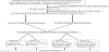
15 subjects do not respect the inclusion criteria and 4 patients were excluded for absence of written consent. Therefore 84 patients (47 men, 37 women), mean age 50±4.7, meeting the inclusion criteria, were evaluated and treated after acceptance of informed consent (figure 1).
All patients were suffering from diabetes and they went to our attention with the diagnosis of diabetic foot syndrome. They were divided into 2 groups (A and B) of 42 subjects according to the absence or presence of associated lymphedema, diagnosed by clinical examination and duplex sonography. Each group was then further divided into two subgroups through a stratified randomization with regard to 3 prognostic factors, smoking status, degree of ulceration, degree of lymphedema in order to have homogeneous groups. Each one made of 21 subjectswho made ESWT at different weekly frequency. All subjects were treated with Dermagoldelectroidraulic device equipped with an unfocused probe (MTS Europe GmbH, Constance, Germany).
Patients in group A-Ireceived 1 session per week of ESWT for 5 weeks (1000 pulses were administered for each session), yet thegroup A-II received 2 session per week (every 84 hours) of ESWT for 5 weeks (500 pulses for each session).
Subjects in groups B, affected by lymphedema, received: 1 session per week of ESWT for 5 week (1000 pulses for each session) for group B-I and 2 sessions per week (every 84 hours) of ESWT for 5 weeks (500 pulses for each session) for group B-II; manual lymphatic drainage was also performed in bothsubgroupsB according to their underlying disease.
The mean energy density applied for each pulse was 0.13mJ/mm2 in all groups.
Before and after treatment period ulcers were classified according to: localization, width (in cm), length (in cm), percentage of granulation tissue, necrotic tissue, fibrous tissue, presence of bacterial exudate (classified as absent, minimal, moderate, high) and pain, which have been assessed by VAS (Visual Analogic Scale). Lymphedema was evaluated by ankle circumference at T0 and T1.
The morphological evolution was monitored taking photos before each session using digital cameras with resolution higher than 5 megapixels and macros function; all pictures were taken at the shortest distance necessary to frame the lesion and the standardized squared. After each session a medication with povidone-iodine, Amukine MED 0.05% and application of semipermeable occlusive dressing was performed; this protocol was identically in all groups and in every session.
No compressive bandage was applied to the patients during the treatment period and was also monitored the possible occurrence of side effects such as pain, petechiae and cutaneous adverse reactions related to ESWT therapy. Lymphedema in groups B-I and B-II was evaluated at T0 and T1 by measuring the ankle circumference with a standard tape measure (MediGmbh, Bayreuth,Germany) [12].
All data are given as means ± SDs. Differences between mean values before and after the rehabilitation period were tested for significance using Student’s t-test for paired observations. The minimum level of statistical significance was set at p<0.05.
3. Results
Before treatment (T0) in group A-I the mean wound dimension was 1.87±0.53 cm2 and the mean VAS score for subjective pain was 5.7±1.47; in group A-II the mean wound dimension at the first evaluation (T0) was 1.69±0.52 cm2 and the mean VAS score was 5.1±1.13; in group B-I the mean wound area at T0 was 2.83±0.97cm2 and the mean perceived subjective pain was 5.5±1.54; in group B-II before treatment (T0) the mean wound size was 2.71±0.68 cm2 and the mean VAS score for subjective pain was 5.3±1.40.
At the end of the 5 weeks treatment (T1) we found:
In group A-I a reduction of wound area from 1.87±0.53cm2(T0) to 0.77±0.34 cm2(p<0.01) and a reduction of the perceived subjective pain from 5,7±1.47 (T0) to 1.8±0.72 (p<0.01); In group A-II a reduction of wound size from 1.69±0.52 cm2 (T0) to 0.45±0.13 cm2(p<0.001); a reduction of VAS score from 5,1±1.13 (T0) to 0.5±0.11 (p<0.001);
In group B-I a reduction of wound area from 2.83±0.97 cm2 (T0) to 1.88±0.59 cm2 (p<0.05); a reduction of the perceived pain from 5,5±1.54 (T0) to 2.9±1.31 (p<0.05); In group B-II a reduction of wound dimensions from 2.71±0.77 cm2 (T0) to 1.54±0.23 cm2(p<0.01); a reduction of the perceived subjective pain from 5,3±1.40 (T0) to 2.3±0.47 (p<0.001).
We found a significant reduction of wound area and VAS score in all groups (figures 2 and 3).
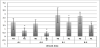
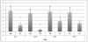
Although there were significant final data in all groups, the best results,in terms of absolute value, were found in groupA-II and B-II, which performed ESWT twice a week with 500 pulses per session. No side effects were found during treatment.
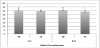
In groups B-I and B-II,whose subjects were affected by lymphedema, we found a reduction of limb circumference. The initial mean circumference of lower limb assessed on the ankle was 30.7±3.1 cmin group B-I and 31.1±2.9 cm in group B-II. Thefinal result was significant only in group B-II which performed the treatment 2 times a week: 3.4 % (final circumference 29.7 cm) in group B-Iand 5.8 %(final circumference 29.3 cm) in group B-II (p<0.05) (figure 4).
4. Discussion
Wound duration is a quantifiable surrogate for one or more undefined variables that can have a profound negative effect on ulcer healing [13]. Has been widely described how the venous system damage, destruction of microlymphatics and local nerve fibers lead to impaired fluid drainage and altered regulatory mechanisms, resulting in impairment of wound healing. Comorbid conditions of diabetes such as obesity, venous insufficiency and rheumatoid arthritis can also interfere with a proper wound-healing response [14].
Several studies showed how ESWT can be able to accelerate wound healing reducing time for cicatrization, promoting the rearrangement of endothelial cells and the basal lamina with a significant increase in the release of growth factors, nitric oxide, TGF-β1 and nuclear proliferation antigen15, 16,by inducing mechanotransduction; they can also reduce the incidence of infections thanks to the bactericidal effect and reduce pain by inducing degeneration of the nerve fibers originating from small neurons ATF3-immunoreactive,which seems to achieve a relief of pain and influence the increase in Substance P [17,18].
According to Stieger [10], which showed an effect of ESWT on limphatic drainage in non-healing chronic leg ulcers, we applied the unfocused Shock Wave treatment on chronic leg ulcers even in case of lymphedema due to the specific wound. Stieger results could be linked with the cavitation effect of ESWT on tissues since the reflection wave induced by the device triggers a jet stream microcavitation resulting in increased tissue perfusion (as shown by Wang et al. [9] in a rat model), reduction of the local inflammatory reaction with upregulation of cell proliferation, particularly of fibroblasts and keratinocytes, and angiogenesis, through an increased expression of VEGF ("Vascular Endothelial Growth Factor")and eNOS (endothelial Nitric Oxide Synthase).
Recently, regeneration of lymphatic vessels has been demonstrated by using physical treatment methods such as extracorporeal shockwave therapy (ESWT) [19], EWST leads to increased cell permeability and expression of growth factors, such as VEGF-C, which is associated with promoting lymphangiogenesis and accelerating capillary morphogenesis [20,21]; Kim et al. used mice suffering from lymphedema at the mid-thigh to investigate the effect of gelatine hydrogels containing VEGF-C and/or ESWT. The combination of treatment methods (VEGF-C hydrogels and ESWT) showed the best results in terms of lymphatic vessel formation, improvement of lymphedema, and enhanced expression of VEGF-C and VEGFR-3 [22].
ESWT increases the platelet–cell adhesion molecule-1 (PECAM-1) production on leukocytes and on endothelial cells which mediate a physical link between the cell surface and the nuclear envelope. It is critically involved in the trans-endothelial migration processes at inflammatory sites, endothelial cell migration, and formation of new blood vessels.This may transmit mechanical or biochemical signals that may modulate the genic expression of cells [23,24].
Schaden et al. [25] and Mariotto et al. [26] demonstrated that ESWT can promote angiogenesis, decrease neutrophils and inflammation, and decrease the number of adipocytes.
Wang et al., 2014 [27] evaluated the long-term effects of ESWT in chronic foot in a long follow up during 5 years. The evaluations included clinical assessment of the ulcer status, including the size, shape, and depth with photo documentation, local blood flow perfusion scan, and the mortality and morbidity (including amputation) in 1 and 5 y after ESWT. Tissue viability was evaluated by local blood flow perfusion scan pre-operatively and at 6 wk, 1 y, and 5 y post-operatively. After ESWT, the blood perfusion rates is greatly improved in both DM (p = 0.011) and non-DM (p = 0.033) groups. The flow perfusion rate was better after 6 weeks, and the improvement was maintained for one year. In both groups, the percentage of blood perfusion decreased in the follow up to 5 years. Non-DM group showed a better blood flow perfusion than that of the DM groupat 1 year to 5 years follow up.
The clinical outcomes, mortality, and morbidity were compared with a database control group of 149 patients with diabetic foot ulcers previously treated by the author. The experimental group showed better results than the data with no significant differences respect the control database. Complications such as amputations were higher in the control group at both one year than five years. The mortality rate was 9.4% in historical controls and 0% to 1 year and 24% to 5 years in the experimental ESWT patients. At the conclusion of this study Wang et al concluded that ESWT appears effective in the treatment of chronic diabetic and non diabetic foot ulcers. However, the effects of ESWT significantly decreased from 1to 5 y after treatment.
The study included 38 patients with 40 ulcers in the diabetes mellitus (DM) group and 29 patients with 32 ulcers in the non-diabetes mellitus (non DM) group. All patients received unfocused ESWT Few studies have investigated the timing of application of ESWT.
Wang in 2009 [9] investigated the ideal frequency of administration and the bio-mechanisms operating during ESWT of wounds in diabetic wounds treatment in rats models. Fifty male Wistar rats were divided into five groups. GroupI consisted of non-diabetic control; group II included diabetic control receiving no ESWT; group III included rats that underwent one session of ESWT (ESW-1) on day 3 (800 impulses at 0.09 mJ/mm (2) post-wounding; group IV included rats that underwent two sessions of ESWT (ESW-2) on days 3 and 7; and group V included rats that underwent three sessions of ESWT (ESW-3) on days 3, 7, and 10. The number of pulses (800) was the same for each session. The wound healing was assessed clinically. Blood perfusion scan was performed with laser Doppler. The VEGF, eNOS, and PCNA were analyzed with immunohistochemical stain. The results revealed that the wound size was significantly reduced in the ESWT-treated rats, especially in the ESW-2 and ESW-3 groups, which performed the treatment with a higher frequency during the week, as compared to the control. Blood perfusion was significantly increased after ESWT compared to the controls. Histological findings revealed a significant reduction in the topical pro-inflammatory reaction in the ESWT group as compared to the control. In immunohistochemical stain, significant increases in VEGF, eNOS, and PCNA expressions were observed in the ESWT group, especially in the ESW-2 and ESW-3 groups, as compared to the control. So he concluded that the treatment with an optimal session of ESWT performed twice or three times a week could significantly enhance diabetic wound healing; this effect was strictly associated with the increase in neo-angiogenesis and tissue regeneration, and topical anti-inflammatory response.
Furthermore Bae in 2013 [21] showed how ESWT is an effective modality in the treatment of stage 3 lymphedema after breast cancer treatment, leading to a reduction of the circumference and thickness of arms with lymphedema providing clinically favorable outcome to patients. In his studyESWT was carried out twice a week for two weeks using electromagnetic type and the stimulus was given on the treatment site according to the patient’s tolerance, 2,000 times in one session with an energy of 0.056-0.068 mJ/mm2. Stimulus was applied 1,000 times to the most fibrotic lesion felt by the examiner’s palpation, and the other 1,000 times was applied to other less fibrotic lesion. CDPT (Complex Decongestive Physical Therapy) or pneumatic compression was not successful on these subjects in the past: so he divided subjects in two groups by their opinion. 4 patients were treated by manual lymphatic massage and pneumatic compression with ESWT; other 3 patients were treated with ESWT only. He found a significant reduction in lymphedema volume, limb circumference, and skin fold thickness in all subjects who were treated with four sessions of ESWT, concluding that ESWT can be considered an effective modality in the treatment of stage 3 lymphedema after breast cancer operation, with or without other traditional treatment modalities.
Christ et al. [28] confirmed the improvement of skin elasticity in the treatment of cellulitis and connective tissue weakness by means of ESWT. Patients in their study received treatment for 6 therapy sessions and were followed up for 3 months. It was observed that the network of collagen/elastic fibers in the dermis and subcutaneous became denser and measurably firmer. In the parallel biomechanical examinations, reduced oxidative stress by ESWT was shown by this study by means of increased lipolysis and by the release of toxic aldehydic products of lipid; furthermore they showed the improvement in collagen synthesis and in skin condition. This study also confirmed the decrease of orange pill sign, which was shown in stage 3 lymphedema, and other measurements of skin conditions including skin fold thickness and skin hardness. This may indicate improvement of skin elasticity and connective tissue strength.
Tinazzi et al. [29] applied ESWT to patients affected by systemic sclerosis showing an improvement of VAS and Rodnan skin score for skin wellness and in increased endothelial progenitor cells and circulatory endothelial cells.
According to Wang [9] we administered the same therapy and the same number of pulses in all groups of study, but with different temporal modalities during the week, by splitting in two times (500 every 84 hours instead of 1000 in a single session) the amount of pulses administered in groups A-II and B-II. So 1000 pulses one time a week were administered in groups A-I (chronic wound) and B-I (chronic wound and lymphedema) and 500 pulses two times a week were administered in goups A-II (chronic wound) and B-II (chronic wound and lymphedema). Results showed how ESWT have been significantly able to improve healing in all groups of treatment, but with a greater result as for absolute values in those groups (AII and B-II) which performed the treatment twice a week. In group B-II, which had concomitant lymphedema and performed the treatment twice a week, we also found a significant reduction of ankle circumference.
5. Conclusion
Considering all this evidences, we can state that the ability of ESWT to act on the repair of microvascular damage in chronic wounds induces, on the one hand the activation of the skin repair process and on the other the recovery of adequate venous and lymphatic microcirculation. In addition, ESWT induces an improvement in blood flow and therefore reduces edema associated with chronic wound, even in the absence of compression bandage.
Particularly, in the skin healing mode processes by which cells react to mechanical forces it is crucial in the switch between healing, failure to re-epithelialization and hypertrophic scar. In vitro studies show that the mechanical stimulus induced by ESWT in metabolic processes that involve the production of fibroblasts and keratinocytes is able to lead the process towards the physiological repair of the wound. Mechanical signals received by fibroblasts guide their transformation from a soft substrate with a little adhesion and poorly developed stress fibers into an adherent strongly geometry, that improves keratinocyte differentiation [7].
The intracellular mechanical tension and the conversion of mechanical signals in biomolecular events, related to the expression of specific genes, are responsible for the contractile force and the creation of a good environment for reparative and regenerative processes, especially when it is associated with the presence of lymphedema.
The progressive pathologic evolution, in fact, related to interstitial lymphatic hypertension and the consequent tissue acidosis, hypoxia, accumulation of lymphocytes, of free radicals and other toxic substances, slowing the healing process. Furthermore, low-energy ESWT have been proven to induce therapeutic lymphangiogenesis by up-regulating vascular endothelial growth factor C and basis fibroblast growth factor, and by improving lymphedema. Therefore, the improvement of lymphedema in our study could be the result of lymphangiogenesis. The change in lower limb circumference in this study could be the result of both manual lymphatic drainage and ESWT promoting reduction of inflammatory cells, reconditioning of skin tissue and improvement of lymphatic drainage.
Based on our clinical experiences and researches related to the effect of ESWT on the healing of chronic wounds [6,30] and the effect of the manual drainage in aiding the healing processes [31,32]we believe that the most appropriate treatment time is 2 sessions weekly with total energy of 2640 Mj (which it is the average energy applied in our previous studies and experiences of six years of work on chronic ulcers) divided in two weekly applications spaced from each other by 84 hours (2 x 1320 Mj)rather than in a single administration (1 x 2640 Mj) in order to keep the healing promoting stimulus without a stimulus deflection phase during the week.
This type of application can not be generalized, it could be probably a best choice for chronic ulcers in presence of lymphedema.
The results obtained in subjects with lymphedema, in which the presence of edema represents a delay element for healing, can encourage us in confirming that the healing process is based on angiogenesis, that is very important to promote drainage, but above all to confirm that shock waves are crucial in reactivating and accelerating the healing process of chronic wounds.
Limitations of the study is the absence of a comparative group (with the same standard treatment but without ESWT); further investigation with a larger sample will be necessary to consolidate our results.
Conflicts of Interest
The authors declare that they have no competing interests.