


1. Introduction
Glycation is a general term covers a series of complex and spontaneous reactions among sugars and amino groups of proteins and further interactions with the advanced glycation end-products (AGEs). Extensive functional proteins involved in glycation result in feedback changes of their physical and functional properties and cause subsequent chronic diseases, such as cataract, arteriosclerosis and Alzheimer’s disease [1-3]. Concerns no matter addressed on healthcare in monitoring the status of protein glycation for specified individuals or development of products bearing functions or effectiveness of anti-glycation, development of an accurate, handy and reliable clinic assessment as a potential disease marker is always a critical issue [1,4-6]. Among the protein glycations, human serum albumin (HSA) glycation has attracted the most concerns and interests because HSA is the most abundant protein in the plasma and sensitive to undergo glycation. Albumin is one of the longest known proteins of plasma, bearing a wide spectrum of physiological functions. Thus, it has been commonly targeted and used for investigation of protein glycation. Among those, serum albumin is the general candidate of albumins. In human blood, albumin presents 50% of the normal individual’s plasma protein [6]. BSA, with 583 amino acids and molecular weight of 66.28 KDa [7], has 76% sequence homology and bears a similar ellipsoidal shape as HSA. Both have three homologous domains (I, II and III), which are connected together through helical extensions [1,7,8].
Structural and physicochemical properties characterization of BSA and HSA have been intensively investigated [1,8-10], a main difference for presence of two tryptophan residues (Trp-131 and Trp- 214) in BSA, while one (Trp-214) is present in HSA [11,12]. The Tryp- 214 localized at domain II of HSA is suggested to have a role in its amyloid fibril formation [13]. Even very minor differences are present between BSA and HSA and perform more or less differently on their responsible functions, both share similar structure and exhibit most biochemical activities in general. In addition, available source, high grade of purity and affordable cost of BSA are appreciated by most users. Accordingly, BSA is regarded as a referenced standard protein and widely used in most laboratories. In this study, in addition to SDS-PAGE characterization of BSA peptides being sensitive to glycation, the peptides were subjected to LC/MS/MS analysis for proteomic figuration focused on some specific amino acid segments present in both BSA and HSA. Perspectives addressed on potential use of those unique segments as markers in product development for status surveillance of human health are raised for discussion.
2. Materials & Methods
2.1 Materials
Bovine serum albumin [BSA, 98% purity] and D-(-)fructose were purchased from Sigma-Aldrich Co. (St. Louis, MO). Potassium diphosphate and dipotassium hydrogen phosphate were purchased from Hayashi Pure Chemical Industries Ltd. (Osaka, Japan); 2-mercaptoethanol and sodium dodecyl sulfate [SDS] were purchased from Merck [Darmstadt, Germany]; Acryl/Bis solution, ammonium persulfate [APS], and TEMED were purchased from Amresco Inc. [Solon, OH]; All other chemicals and solvents used in this study were of analytical grade.
2.2 Glycation of BSA and fructose for various intervals
In vitro glycation between BSA and fructose was conducted according to the procedure reported previously [14]. Briefly, BSA was dissolved in 0.2 M phosphate buffered-saline (pH 7.4, containing 0.06% sodium azide) to yield a stock solution of 60 mg/mL. This solution was applied to prepare a series of solutions containing fructose prepared in phosphate buffered-saline (pH 7.4) and deionized water to a final concentration of 20 mg BSA/mL and 0.5 M fructose. Reaction mixtures were incubated at 50°C for 0, 2, 4, 6, 8, 12 and 24 h. After incubation, the reacted solutions were stored frozen at -20°C for further analysis.
2.3 Sample preparation for SDS-PAGE analysis
From each tube after glycation reaction, 200 μL reactant was withdrawn and transferred to a centrifugal concentractor (Vivaspin® 2 10,000 MWCO PES tube, GE Healthcare, Amersham, UK) previously deposited with 1.3 mL deionized water. After mixing the tubes were centrifuged (3000 g) at 4oC for 5 min. To each tube, deionized water was replenished to reach the level of 1.5 mL and pipetted for 50 times to thoroughly mix prior to centrifugation. This step was repeated once. Then, deionized water was replenished to reach the level of 1 mL and well mixed by repeated pipetting. From which 100 μL of each solution was withdrawn and well mixed with 100 μL of SDSPAGE sample buffer in an Eppendorf tube. The tubes were capped and heated in a water bath at 100oC for 10 min. After heat treatment, 10 μL of the solution was loaded into a well of a prepared SDSPAGE gel (10% polyacrylamide separation gel containing 0.1% SDS) and subjected to electrophoresis (Mini-Protean 3 System, Bio-Rad, Hercules, CA) run at 100 V for 180 min. Then, the gels were stained with Coomassie Brilliant blue R-250 (Sigma) for 20 min and destained with aqueous acetic acid/methanol solution (100 mL of acetic acid and 200 mL of methanol per liter). The gels were scanned by an image scanner (U9909-H7L0, Amersham Biosciences, Uppsala, Sweden) and densitometric quantification using Fujifilm Multigauge ver. 3.0 Analysis Software (Fujifilm, Tokyo, Japan).
2.4 Proteomic analysis
From the SDS-PAGE, the band with estimated molecular weight (MW) 97 KDa (Figure 1A) was cut from the 24 h-glycation gel lane and the band with MW 50 KDa was cut from the 0 h-glycation gel lane with a scalpel. The gel stripes were subjected to in-gel tryptic digestion, and then applied to Thermo Finnigan LCQ DECA XP Plus Ion Trap Mass Spectrometer for peptides analysis (Instrument Center of National Cheng Kung University, Tainan, Taiwan). Peptides were separated by Zorbax 300 SB-C18 column (150 x 0.075 mm, 3.5 μm, Agilent ) using a mobile phase that contained Solvent A (100% H2O with 0.01% Formic acid) and Solvent B (100% Acetonitrile with 0.01% Formic acid ) at a flow rate of 0.2 μL/min. The mobile phase composition was gradient from 0 to 10% solvent B for 2 min and then changed from 10 to 60% solvent B over 40 min. The mass spectrometer was scanned from 500 to 3000 m/z range. The peptide fragments were searched by MASCOT web (http://www.matrixscience.com) interface from NCBInr 20121013 (21019903 sequences; 7205775305 residues) databases. The coverage ratios were calculated based on the sequences of BSA gi|1351907 and gi|3674602 (NCBInr Database). In further, 299 manual queries for targeted m/z scanning addressed on the most likely glycol-conjugates formed between AGEs and the glycation precursor peptides in the 97 KDa (Table 1) [15,16].
2.5 List of the proteomic search parameters
Type of search : MS/MS Ion Search
Enzyme : Trypsin cuts C-term side of KR unless next residue is P.
Variable modifications : Carbamidomethyl (C), Deamidated (NQ),
Gln->pyro-Glu (N-term Q), Glu->pyro-Glu (N-term E), Oxidation
(M).
Mass values : Monoisotopic
Protein Mass: Unrestricted
Peptide Mass Tolerance : ± 1 Da
Fragment Mass Tolerance: ± 1 Da
Max Missed Cleavages : 2
Instrument type: ESI-TRAP
Number of queries : 299.
3. Results and Discussion
3.1 SDS-PAGE profiles of BSA after reaction with fructose
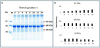
SDS-PAGE spectra of BSA after reaction for glycation with fructose at 50oC for 24 h are shown in Figure 1A. Generally, in reference to the marker proteins (M), in addtion to the major band of BSA located at MW 66 KDa and crosslinking complex proteins visualized at the top of gel margin, bands with MW 97 KDa and 50 KDa were with detected. In comparison to original BSA spectrum (Lane 0), the bands with 97 KDa are apparently the glycation products. Their band intensities increased with time of glycation (Figure 1B). Appearnce and quantification of 97 KDa band is normally regarded as measure of glycation [1,4,14-16]. As noticed, quantities of an original but minor band of BSA with MW 50 KDa decreased gradually with time of glycation reaction (Figures 1A and 1B). Based on band intensities of 66 KDa shown in (Figure 1B), quantity of the major BSA comprising peptides also decreased gradually with time of glycation.
3.2 Proteomic analysis of the 97 and 50 KDa peptides
After LC/MS/MS analyses, the trypsin-digested peptide sequences were subjected to MASCOT search with NCBInr Database for fingerprint matching with BSA gi|1351907 (Figure 2). For the 97 KDa peptide, the search results are: Score: 1634, Matches: 69(30), Sequences: 29(18), Protein sequence coverage 49%. For the peptide with MW 50 KDa, the results are Score: 1392, Matches: 74(28), Sequences: 26(11), Protein sequence coverage 42%. Accordingly, it is undoubted that 97 and 50 KDa peptides are originating from BSA family. The BSA (RecName: Full=Serum albumin; AltName: Full=BSA; AltName: Allergen=Bos d 6; Flags: Precursor) comprises 607 amino acid residues with MW 69.2 KDa and 5.82 of isoelectric point (pI). When MASCOT search was applied to fingerprinting match with another NCBInr database of BSA gi|3674602, the coverage ratios for 97 and 50 KDa were 51 and 44%, respectively. The polypeptide of gi|367460260 (Chain A, Crystal Structure of Bovine Serum Albumin) contains 583 amino acid residues with MW 66.2 KDa and 5.60 of pI. In comparison, both referenced polypeptides are only different in the first 24 aa. Basically, BSA gi|3674602 is a mature protein translated and transported after deletion of the first 24 amino acids observed in BSA gi|1351907. In this study, detailed searches are mainly based on BSA gi|1351907 (Figure 2).
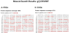
In further comparison of the matching peptide segments between 97 and 50 KDa polypeptides, many commonly overlapped segments are observed (marked with red color shown in Figure 2). In BSA domain 3 (aa 375-607) [1,4], the matched segments in 97 and 50 KDa peptides are identical except that K548 is only matched in 50 KDa peptide. However, there are 5 segments, belonging to BSA domains 1 and 2, only matched in peptide of 97 KDa. It is obvious that 50 KDa is located in BSA domain 3. In literature, the lysine residues including K256, K299, K420, K439 and K548 are regarded as active amino acid residues involved in glycation [1,8,15-17]. When BSA was subjected to reaction with glucose or galactose at 60oC for 120 min, K256 and K420 have been noticed as sensitive residues involved in glycation [16]. As generalized, those lysine residues except K548 (only detected in the peptide of 50 KDa) have been matched in both polypeptides. Thus, K548 might have been modified during glycation and, thus, not detected in 97 KDa.
Among the comparisons, it is of interest to point out the difference that the 10 aa-segment between E300 and K309 and K548 are only matched in the 50 KDa. This segment of ECCDKPLLEK (aa 300- 309) has been demonstrated as a glycation precursor by Ahmad et al. [15]. In that study, BSA has been subjected to reaction with glucose at 37oC for 5 weeks, the ECCDKPLLEK segment changed after 2 week reaction and disappeared after 5 weeks of reaction. Based on the finding that the SDS-PAGE detected 50 KDa containing this glycation sensitive 10 aa-segment but not involved in formation of the glycated 97 KDa peptide (a commonly detected indicator of glycated protein), decrease of 50 KDa provides an alternative approach to monitor status of blood glycation.
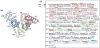
As a further comparison addressed on the sequences adjacent to this 10 aa-segment between BSA and HSA (human serum albumin) (Appendix Figure 1) [8], KPLLEK in this segment along with the followed 8 aa-segment of SHCIAEVE are identical in both albumins. Therefore, the 14 aa-segment in HSA could be regarded as glycation sensitive target and used to develop a surveillance to monitor status of human blood. Similarly, in consideration of K548 as an active lysine residue in 50 KDa, the adjacent 11 aa-segment of Q545-L555 (QIKKQTALVEL) (Appendix Figure 1) is also identical between BSA and HSA and can be used as a target to develop a measure in monitoring human blood glycation as well.
3.3 Queries in detection of AGEs-glycation precursor formation
Segments of glyco-conjugates formed between AGEs and the glycation precursors have been searched by queries based on m/z values obtained from LC ESI MS/MS (Table 1). As expected, K420, K557 and K580 are sensitive to react with fructose to form Schiff base/ AP as AGEs-precursor conjugates. K420 and K557 are also sensitive to form CML, pyrraline, imidazolon and AFGP as glyco-conjugates [15-20]. In addition, K214, R507, K523 and K526 have also been noticed being involved in formation of AGEs-precursor conjugates detected in the peptide of 97 KDa. As reported by Ahmad et al [15] to incubate BSA and glucose at 37oC for 5 weeks, conjugates of H402/ K412+Schiff base/AP was detected after 2 weeks of reaction and not detected after 5 weeks of glycation. This means that, after a prolonged period of glycation, the AGEs-precursor conjugates might degrade or be further modified.
When a query was made addressed on the 10 aa-segment (E300-K309, which was not matched in 97 KDa) as a precursor, the suspected AGEs-precursor conjugates was not detected (Table 1). This is further supporting that glycation of 50 KDa was not involved in 97 KDa formation. As queries were made addressed on K548, which was matched in the peptide of 50 KDa (Figure 2), it is noteworthy to point the finding that that K548 is sensitive to react with Schiff base/ AP, CML, imidazolon and AFGP (Table 1) [15]. This is an evident clue to support why K548 was not matched in the glycated peptide of 97 KDa. Direct determination of AGEs and characterization of the AGEs-precursor conjugates to identify their possible pathophysiological roles for various diseases have been intensively investigated [1,4,5,17,20-22] As mentioned above that the adjacent 11 aa-segment of Q545-L555 (QIKKQTALVEL) was similar to that if HSA (Appendix Figure 1), this segment deserves to be further used as a target to develop a measure in monitoring human blood glycation.
4. Conclusion
After subjection of BSA to an in vitro glycation with fructose at 50oC for 24 h and followed by SDS-PAGE analysis, an original 50 KDa peptide was disappearing with time along with appearance of the normally detected 97 KDa glycated peptide. When the 50 KDa and 97 KDa bands were cut and subjected to LC MS/MS proteomic analysis and MASCOT fingerprint matches with sequences of BSA gi|1351907 and BSA gi|3674602 (NCBInr Database), the coverage ratios were 42 and 44% for 50 KDa and 49 and 51% for 97 KDa, respectively. The segment of ECCDKPLLEK (aa 300-309 sharing a common segment of KPLLEK with that in HSA) was only detected in the 50 KDa peptide. Apparently, 50 KDa peptide is glycation sensitive but may not involve in formation of the detected glycated 97 KDa peptide. In further search by manual queries addressed on m/z values of the AGEs-precursor conjugates addressed on specified segments, K548 was of particular detected being active to form various AGEsprecursor conjugates. The adjacent 11 aa-segment of aa 545-555 (QIKKQTALVEL) is identical to that of HSA and deserves to use as a target to develop a measure in monitoring human blood glycation. Thus, segments of KPLLEK (aa 304-309) and QIKKQTALVEL (aa 545-555 containing K548 are of potency to use as a glycation-sensitive marker for development of rapid clinic assessment in surveillance of human health. In further, the segment of ECCDKPLLEK (aa 300- 309), has been demonstrated in the literature as a glycation precursor, was exclusively detected in the 50 KDa peptide. This peptide is of potency to be used as a potential marker in development of a clinic alternative to measure status of human blood glycation.
Competing Interests
The authors declare that they have no competing interests.