


1. Introduction
Neonates staying in the Neonatal Intensive Care Unit (NICU) frequently receive procedures accompanied by pain for examination and therapy. Pain that neonates receive in the period of complex and rapid brain development have short- and long-term negative influences on the development of the brain and nerves and cognition, motion, and behavior of neonates. Heel lance is a blood sampling procedure and it is the most frequent and common painful procedure applied to neonates staying in the NICU. Therefore, the development of pain relief for the pain of neonatal heel lance has been attempted for a long time, but no conclusion has been reached with regard to safe and effective pain relief for neonatal heel lance. One of its reasons is problems with pain evaluation due to a lack of pain expression with words by neonates. In this report, trends of pain evaluation indexes used in neonates and the relief of the pain of neonatal heel lance are discussed and then benefits and future prospects of evoked potentials (EP) recently attracting attention as a neonatal pain evaluation index are introduced.
2. Subsequent Influence of Pain in Neonates
Pain detected by peripheral nociceptors is transmitted to the cerebral cortex via the spinal cord, brainstem, and thalamus [1,2] . At the same time, pain transmission from the spinal cord to the brain is inhibited by descending inhibitory controls, adjusting pain [1]. Development of this pain transmission and pain modulation rapidly starts at 22 weeks of gestation and mature 2 months after birth in healthy full-term infants [1-5].
Neonates staying in the NICU, especially preterm infants born earlier than 37 weeks of gestation, receive painful procedures 7.5- 17.3 times a day for examination and treatment in this complex and rapid developmental period of the pain transmission pathway [6,7] , so that preterm infants are exposed to the risks for poor early neurodevelopment, developmental disorder of the brain, and delay in postnatal growth [1,8] . The nerve activity level is high during heel lance in preterm infants who received intensive care for at least 40 days or special treatment compared with that in age-matched term infants who did not stay in the NICU [9]. In addition, in a cohort study involving preterm infants, frequent pain was associated with lower body weight and head circumference at 32 weeks [10] and a reduction of the white matter and subcortical gray matter at the termequivalent age [11] even though other medical confounding factors were adjusted.
Repeated pain in the neonatal period has a long-term negative influence. In preterm infants, poorer cognition and motor function in infancy [12], a reduction of Full Scale IQ (FSIQ) at school age [13], and a reduction of the cognitive score in late adolescence [14] have been reported even though other medical confounding factors were adjusted for (after comprehensively adjusting for multiple clinical factors). Moreover, internalizing behaviors, such as depression and anxiety, were clearly noted in infancy in preterm infants compared with full-term infants even though other medical confounding factors were adjusted for [15], and it continues to school age, late adolescence, and young adulthood [16-20].
Painful procedures in the NICU include heel lance, blood sampling, arterial puncture, lumbar puncture, and placement and intramuscular injection, and heel lance is one of the most frequently performed painful procedures [21]. Of the painful procedures performed 3,605 times in total in 55 neonates who stayed in the NICU for 28 days or longer, heel lance accounted for 71% [22]. However, the pain of the most frequently performed heel lance in the NICU is not sufficiently managed. In Italy, 30% of 140 medical workers of 5 NICUs (89 nurses and 51 physicians) answered that no intervention is performed for heel lance [23].
Neonates staying in the NICU cannot avoid frequent pain stimulation for treatment and examination. To prevent a negative influence on the development of the brain and nerves and cognition, motion, and behavior, it is essential to manage the pain of heel lance most frequently performed in the NICU.
3. Pain Evaluation Index for Neonates
For the neonatal pain assessment tool, a mixture of physiological and behavioral indices has been recommended [24,25] . Many pain evaluation indices are prepared by comprehensively mixing facial expressions (grimace), physiologic measurements (vital signs, such as heart rate and blood pressure, respiratory rate, and pulse-oximetry readings/oxygen requirement), and behavioral components (crying/ consolability or motor activity) [24].
For the pain assessment tool for acute and procedural pain in children including neonates, Premature Infant Pain Profile (PIPP) and Premature Infant Pain Profile-Revised (PIPP-R), Neonatal Infant Pain Scale (NIPS), Crying, Requires oxygen, Increased vital signs, Expression, and Sleeplessness (CRIES) (neonatal period), Distress Scale for Ventilated Newborn Infants (DSVNI) (Ventilated neonates and infants), Douleur Aigue du Nouveau-né (DAN) (neonatal period-3 months), Neonatal Facial Coding System (NFCS) (neonatal period-18 months), and Evaluation Enfant Douleur (EVENDOL) (neonatal period-6 years old) have been reported [26,27] .
In many studies on pain relief for neonatal heel lance, PIPP or PIPP-R is selected for the pain evaluation index [28,29] . PIPP is comprised of 2 physiological indices (heart rate and oxygen saturation) and 3 behavior indices (brow bulge, eye squeeze, and nasolabial furrow) and the score is corrected with the gestational age and behavioral state [30]. The score varies from 0 to 21, a score of 6 or lower represents that there is almost or completely no pain, and a score of 12 or higher represents moderate to severe pain [31]. The reliability and validity of PIPP have been demonstrated [30,32,33]. In PIPP-R, weighting by the gestational age and behavioral state in PIPP was modified [34,35] and between-rater reliability [34] and construct validity [34,35] have been demonstrated.
4. Study on Pain Relief for Heel Lance
4.1 Insufficiency of analgesic effect and adverse effect of pharmacological intervention
It has been reported that no pharmacological intervention exhibited an analgesic effect for neonatal heel lance but adverse effects developed.
In a study on heel lance involving 42 neonates, no significant difference was noted in DAN between the morphine and placebo groups (5% glucose dextrose infusions) [36]. In a study on heel lance involving 30 neonates, morphine did not exhibit an analgesic effect on PIPP-R and morphine induced a reduction of oxygen saturation and prolongation of bradycardia time [37].
In a study on heel lance involving 72 neonates, there was no significant difference in PIPP between the paracetamol and placebo (cherry elixir) groups [38]. In a study on heel lance involving 75 neonates, no difference was noted in the facial action or cry scores between the paracetamol and placebo (sterile water) groups, showing no analgesic effect [39].
In a study on heel lance involving 106 neonates, no significant difference was noted in PIPP between the Lidocaine-Prilocaine (EMLA) cream and placebo cream (Glaxal) groups [40]. Similarly, no difference was noted in pain cry between the Lidocaine-Prilocaine (EMLA) cream and placebo cream groups, showing no analgesic effect, in a study on heel lance involving 112 neonates [41]. In 8 studies investigating the analgesic effect of EMLA cream on invasive treatment including heel lance in neonates, no sufficient evidence for the efficacy of the analgesic was acquired because of insufficiency of the sample size and diversity of the measurement index [42].
Accordingly, because of the lack of the analgesic effect and safety of pharmacological intervention, development of pain relief by non-pharmacological intervention for neonatal heel lance has been promoted.
4.1 Achievements and problems of non-pharmacological intervention
The PIPP scores of oral sucrose (2.74-5.0) [29,43,44], combination of pacifiers and oral sucrose (3.0-5.7) [29,45-47], and combination of sucrose and music (3.0) [44] for the pain of neonatal heel lance were 6 or lower, but an increase in oxidative stress [48] and delay in nerve development [49,50] induced by frequent administration of sucrose contained in all intervention methods have been reported.
For the pain of neonatal heel lance, the effect of pain relief by the interventions described below and combination thereof has been investigated, but the PIPP score was 6 or higher in all conditions, remaining pain: pacifiers (6.4-9.5) [45-47,51-55], facilitated tucking (3.8-9.6) [51,52,54,56-59], breast milk (3.0-9.7) [52,60-62], swaddling (7.0-10.7) [63,64] , kangaroo care (4.1-8.9) [64-67], and holding (13.3) [61].
The PIPP scores of a combination of music, pacifiers, facilitated tucking, and holding [68] and a combination of music and facilitated tucking [69] for pain of neonatal heel lance were 6 or lower (3.6-5.1) and no adverse effect was reported.
Due to the lack of the analgesic effect and safety of pharmacological intervention and adverse effect of oral sucrose, the methods showing an analgesic action on the pain of neonatal heel lance are a combination of music, pacifiers, facilitated tucking, and holding and a combination of music and facilitated tucking. At present, the effective and safe pain relief for neonatal heel lance is being searched for in diverse combinations of interventions.
5. Appearance of Evoked Potential (EP) and Limit of Premature Infant Pain Profile (PIPP)
The potentials evoked by pain represent cerebral cortical activity induced by pain stimulation transmitted through the peripheral nociceptor, spinal cord, brainstem, and thalamus [1,2]. The 2 main types of pain sensing neurons in the skin and other peripheral tissues are myelinated Aδ and unmyelinated C nociceptor [70-72]. Aδ fibers respond to rapid, pricking, and localized pain and C fibers respond to diffuse, burning or aching sensation [70,73-76]. Selective stimulation of these fibers and recording EP have recently become possible. Intraepidermal electrical stimulation is a method to selectively stimulate Aδ fibers.
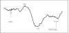
We previously reported that EP consisted of 226.6±8.2 ms negative waves (N2) and 328.2±10.9 ms positive waves (P2) by intraepidermal electrical stimulation in 18 adults (Figure1) [77], showing selective activation of Aδ fibers. The values of EP parameters recorded, such as the latency of N2 and P2, amplitudes of EP (N2-P2), and sensory threshold, were within the range of the results of our and another group’s studies, which employed similar common parameters [78-82].
On the other hand, nerve transmission of pain stimulation is delayed in neonates because myelination is lacking and synaptic currents are immature [83,84] . The latency of the EP by heel lance in neonates (403-420 ms negative wave (N2) and 413-538 ms positive wave (P2)) is longer than that by the stimulation of Aδ fibers in adults [85-88], and the activation of Aδ fibers by heel lance in neonates has not been clarified. However, in neonates, when compared with painless tactile stimulation, the amplitude of heel lance-EP was significantly higher, showing that the pain of heel lance is present by the EP [85-88].
Heel lance-EP pointed out the limit of pain evaluation using PIPP. In a study in which low-intensity pin prick stimulation was applied to 30 full-term infants, the EP was significantly higher than that in the background period, but no difference was noted in PIPP [88], suggesting that the detection sensitivity of PIPP is insufficient for low-intensity noxious stimulation.
Moreover, the indication of PIPP is limited to the neonatal period [30]. The pain evaluation index has to be changed with child growth, such as Face Legs Activity Cry Consolability (FLACC) [89] in infancy-childhood and Face Scale [90] in childhood and thereafter. Accordingly, continuous pain evaluation with growth is difficult for many pain evaluation indices.
Therefore, pain evaluation using PIPP alone is not sufficient. Electrophysiological EP more sharply detecting pain stimulation are expected to contribute to the development of pain relief for neonates [91].
6. Development of EP
To develop pain relief for heel lance for neonates unable to express pain with words, EP may serve as an index to evaluate the pain relief effect. Regarding the action of sucrose in the neonatal heel lance procedure, the PIPP score was significantly lower in the sucrose (n=29) than sterile water group (n=30), but there was no difference in the EP between the 2 groups [86]. This finding suggested non-effectivity of sucrose for the pain of neonatal heel lance [86]. In addition, in a study on the pain relief effect of slow tactile stimulation in neonatal heel lance, the EP after heel lance in neonates who received slow tactile stimulation was significantly smaller than that in neonates after heel lance in the control group (no touch) [92]. Regarding the EP, the pain relief effect of slow tactile stimulation during neonatal heel lance has been reported [92].
In addition, EP-based pain evaluation may enable continuous pain evaluation along with growth. Pain stimulation-induced EP have reported in the neonatal period, infancy at one year old, and at school age and thereafter. Studies on EP-based pain evaluation were performed in neonates at 35 weeks of gestation to 45 weeks of postmenstrual age at which distinguishing tactile and pain sensations is established [85-88]. Furthermore, EP similar to those induced by neonatal heel lance have been detected in a study on vaccination of 17 infants [93]. In a study in which laser stimulation was applied to healthy subjects in childhood and thereafter (7-72 years old, 237 subjects), negative and positive waves, which are pain-induced EP, were clearly detected in all subjects [94]. EP-based pain evaluation may enable continuous evaluation of neonatal pain along with growth and development.
7. Conclusion
Pain evaluation using PIPP is frequently employed in neonatal heel lance, but PIPP has a limit of pain detection sensitivity. EP capable of sensing low-intensity pain without judgment by a rater may serve as an index capable of evaluating pain objectively and quantitatively for neonates unable to express pain with words. The development of EP-based pain evaluation promotes the development of pain relief for neonatal heel lance.
Competing Interests
The author declare that there is no competing interests regarding the publication of this article.
Author’s Contributions
Yui Shiroshita were involved in all the process of this study from the conception of this study to drafting and final approval of the manuscript. Hikari Kirimoto, Kei Nakagawa, Hiroko Uematsu contributed to the writing and final approval of the manuscript. Ikuko Sobue contributed to the conception and design of the study.