


1. Introduction
Options for surgical management of rotator cuff disease include: subacromial decompression and bursectomy, debridement of partial tears and surgical repair of partial or full thickness tears. The standard technique for a repairable rotator cuff tear involves suture anchor placement into an osseous bed prepared in the humeral tuberosity, with a variety of different options for suture configuration. Multiple anchors may be required for acceptable repair.
While many factors influence the outcome of rotator cuff surgery, the most common complication is structural failure of the tendon repair [1]. Better patient-reported outcomes have been shown in patients with structurally intact repairs [2,3], with an improved range of motion and strength compared to patients with structural retears [4-6]. The weakest region of the tendon repair is the suture-tendon interface, which typically becomes a problem in the case of pullout of the suture through the tendon [7]. This complication prompted the senior author to explore single-row suture techniques, which provide a large footprint contact area for the bone tendon interface, as well as creating adequate contact pressure between the suture and tendon. Clinical experience has shown that this technique is amenable to healing and not prone to cutting out. In vitro studies also support the biomechanical efficacy of this technique [8,11].
The section below presents an intra-operative description of this technique.
2. Indications
Arthroscopic or mini-open evaluation of rotator cuff tears deemed to be appropriate for repair with the use of suture anchors.
3. Technique
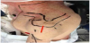
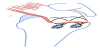
Mini-open surgery is performed under general anaesthesia with optional regional nerve blocks. The patient is placed in the lateral decubitus position with the operative arm placed in traction – typically 10-15lbs (4.5-7kg) (Figure 1). Appropriate antiseptic preparation and draping is performed. The patient receives prophylactic intravenous antibiotic before the skin incision. A straight longitudinal incision measuring approximately 3-4cm in length is placed at the midportion of the lateral acromion. A deltoid split approach is utilised to present the rotator cuff tear for assessment and treatment, typically after arthroscopic acromioplasty and bursectomy.
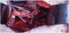
Sutures are placed into the opposing torn ends of the rotator cuff defect using No.1 Vicryl; this will enable manual retraction and increase manoeuvrability of the torn tendons. A Cobb elevator is used to free adhesions on the bursal and articular sides of the rotator cuff, minimizing tension in the repaired tendon. The surface of the tuberosity is decorticated with the aid of a low speed (8000rpm) burr exposing a cancellous footprint. A suture anchor with two loaded sutures (four strands) is selected. The author prefers non-absorbable suture anchors with No.2 Fiber Wire or Ethibond braided sutures. The anchor awl is introduced into the central midpoint of the footprint at Deadman’s angle, as described by Burkhart [9].
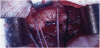
The cruciate suture repair consists of two overlapping stitches, oriented at 45° to the tendon, acting to increase tendon-bone contact and prevent tendon slippage. With the use of a mayo needle, the single-row cruciate stitch is placed using an opposing near-far stitch. The first stitch is placed far (12mm from the torn tendon edge). The second limb of this stitch is placed into the opposite tendon edge in the near position (4-5mm from the torn edge seen in Figure 2). The technique is then repeated with the second suture from the anchor, the tendon can now be laid flat on the bone with the assistance of the tacking sutures (Figure 3). The sutures from the anchor are now tied creating a cruciate stich (Figures 4 and 5). The initial knot tied for each stitch is a sliding half-hitch, this is locked by three half-hitch knots tied at alternating posts. Knots are positioned immediately above the repaired tendon. If the repair requires, two side by side single row cruciate sutures can be placed (Figure 6).

Standard closure techniques are performed and post operatively the patient is placed into a broad arm sling. Typically, this operation can be performed as a day or overnight stay procedure. Pendulum activities are encouraged for the first 4-6 weeks, followed by a graduated return to function and physiotherapy-led rehabilitation program.
4. Discussion
This paper outlines the single-row cruciate suture technique. Traditional single-row suture configurations can pass through poor quality tendon edges, thereby potentially adding to adverse tendon suture pull-out. The cruciate technique, however, passes through both good quality tendon (far stitch) for increased pullout strength, and marginal tendon (near stitch) for apposition. While double-row techniques may provide superior contact area and ultimate tensile strength compared to traditional single-row repairs [10], they are associated with higher material expenses and prolonged operating time and are technically more challenging for the surgeon.
The single-row cruciate suture technique has been validated in vitro and published by the lead surgeon (MR) [8]. In an animal study utilising infraspinatus tendons from lamb’s shoulders, the cruciate suture repair established a significantly greater footprint contact area compared to the Mason-Allen repair (mean difference 101 mm2, p = 0.003). The average footprint contact pressure of the cruciate suture repair (0.78 MPa) was similar to that of the Mason-Allen (0.74 MPa) and double-row repairs (0.79 MPa). The ultimate tensile strength of the cruciate suture repair was significantly greater than that of the transosseous repair (mean difference 62.4 N, p = 0.002).
The authors therefore concluded that cruciate suture repair may improve strength and healing at the repaired tendon rotator cuff insertion compared to other single-row repair techniques. Further animal studies have demonstrated Gap formation in the single-row cruciate suture repair is significantly lower than that of the Mason- Allen repair (mean difference = 0.6 mm, P = 0.009) and no different from that of the Suture Bridge repair (P > 0.05) at 200 cycles [11].
5. Conclusion
Compared to double-row techniques, single-row procedures can utilise half the number of suture anchors, adding to its cost efficacy and promoting sustainable resource management. Furthermore, the marginal difference in mechanical performance of the double-row repair has not been definitively shown to directly translate to superior clinical performance [12,13]. In contrast, the single-row cruciate repair technique described in this paper is a simple configuration, which is easy to master with biomechanical properties validated by in vitro testing [8,11].
Competing Interests
The authors declare that they have no competing interests.