


We have accumulated experimental evidences that a nuclear protein, histone H1, and its immunosuppressive autoantibody (Ab) are associated with post-transplant rejection and tolerance following liver transplantation [1-6]. In clinical setting, we have observed that serum level of histone H1 increased during rejection after liver transplantation while anti-histone H1 Ab was detected in liver transplant patients whose treatment with immunosuppressive drugs was withdrawn or reduced [7].
Protein moonlighting (or gene sharing) is a phenomenon by which a protein can perform more than one function [8] The detection of a protein in unexpected locations within cells, cell types, or tissues may suggest that a protein has a moonlighting function. In the nucleus, the original function of histone H1 has been well known as a DNA binding protein which binds to linker DNA between two nucleosomes and involves in the formation of higher order chromatin structure [9]. However, recent findings clearly demonstrated that extracellular histone H1 have completely different functions as so called, one of the “moonlight” proteins. Indeed, histone H1 is listed at two databases of moonlighting and multitasking proteins (http://wallace.uab.es/multitask/ and http://www.moonlightingproteins.org/ ). Beyond the wall of nucleus, histone H1 translocates to mitochondria and acts as a mediator for apoptotic signal [10]. Furthermore, histone H1 expresses on the cell-surface of macrophages and acts as a receptor for thyroglobulin [11]. We and others found the extracellular nuclear proteins including histone H1 and high-mobility group box 1 (HMGB1) modulate immune responses in the course of immune cell activation and inflammation [12-31]. The trap of extracellular nuclear proteins after surgery may prevent not only rejection but also postoperative sepsis and disseminated intravascular coagulation (DIC) [20,21,28,29,32].
As for immunological function of histone H1 as a moonlight protein, we have demonstrated that the maturation of dendritic cells (DCs) and subsequent T cell activation are enhanced by histone H1 [12]. Another nuclear protein, high-mobility group box 1 (HMGB1), is also involved in mechanisms of DC maturation [13]. Under a certain circumstance, nuclear proteins are secreted from nucleus of “dead” cells by necrosis, apoptosis or autophagy to systemic blood flow. These secreted nuclear proteins are currently being paid attention as damage-associated molecular patterns (DAMPs), signal/mediator or alarmins for cell death to spread inflammation in disseminated intravascular coagulation (DIC) and subsequently induce multiple organ failure (MOF) [22,25]. These results suggested that the detection of extracellular nuclear proteins has a possibility to be a novel diagnostic marker for sepsis, DIC or MOF after surgery as well as transplantation-related rejection. Recently, we have developed a kit to detect circulating histone H1, and are currently evaluating its sensitivity and specificity for post-transplant liver allograft rejection in experimental (Figure 1) and clinical settings.
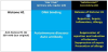
Regarding therapeutic application of anti-nuclear Ab in surgical field, our previous studies have demonstrated that either treatment of recipient rats with commercially available anti-histone H1 polyclonal Ab or immunization with calf thymus histone H1 could suppress acute rejection and prolong allograft survival in a rat heterotopic heart transplantation model [1,34]. We have also reported that the induction of autoimmune hepatitis, which produces anti-nuclear Abs, could overcome acute rejection and prolong liver allograft survival in a rat model of acute rejection after orthotopic liver transplantation (OLT) [16]. Immunologically, the blockade of histone H1 modulated DCs toward tolerogenic status, decreased the cytotoxicity of lymphokine activated killer and natural killer cells, and induced CD4+CD25+ regulatory T cells [2,12]. For further analysis of this mechanism, we generated an immunosuppressive monoclonal Ab against histone H1 (clone: 16G9) and determined a short peptide fragment, SSVLYGGPPSAA (SSV), that binds directly to 16G9 mAb (341). The binding of SSV to 16G9 mAb or serum of both tolerogeneic OLT rats and clinical drug-free OLT patients was inhibited by histone H1. Furthermore, SSV mAb or immunization of mice with SSV induced immunosuppression in serum, suggesting that SSV was an epitope responsible for the immunosuppressive activity of 16G9 mAb [34]. Moonlighting function of histone h1 and its antibody was summarized in Table 1. A novel insight to the role of moonlighting nuclear proteins will allow us to establish a novel diagnostic and therapeutic strategy in post- operative complications as well as liver transplantation.
Competing Interests
The authors declare that they have no competing interests.
Author Contributions
Conceived and designed the experiments: TN,SG,SK, CLC.
Performed the experiments: TN, KCC, LWH, YC.
Analyzed the data: TN, SG, TG, SS, NO.
Contributed reagents/materials/analysis tools: YT, YM