


1. NF1 is a Tumor Disease with Tumor-independent Multi- Organ Involvement
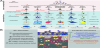
Neurofibromatosis type 1 (NF1) is a tumor disorder (MIM#162200) that is caused by autosomal dominant loss-of-function mutations in the NF1 gene encoding the RAS_GTPase-Activating Protein (Ras-GAP) neurofibromin [1]. Mutation or loss-of-function of neurofibromin leads to hyperactive RAS protein and the subsequent downstream MAPK/MEK signaling pathway [1,2]. Activation of RAS/MAPK/ MEK signaling affects in a cell dependent manner PI3K-mTOR, c-Jun N-terminal kinase (JNK), JAK-STAT3 signaling, cAMP-PKA, Rho- ROCK-LIMK2-Cofilin, or Rac1-Pak1-LIMK1-Cofilin pathways [3].
Constitutional NF1 mutations cause the tumor susceptibility in NF1 patients. Most frequently, NF1 patients are affected by cutaneous, benign neurofibromas, which are composed of Schwann cells, other nerve cells, fibroblasts, mast cells, and endothelial cells [4]. Plexiform tumors, which are another group of frequent tumors in NF1, represent a severe life risk for NF1 patients after transformation into malignant peripheral nerve sheath tumors (MPNST) [5,6].
Although primarily identified as a tumor entity NF1 patients present with multi-organ involvement affecting a broad range of tissues such as skin, skeleton, cardiovascular system, nervous system, and ophthalmic system [7,8]. Although not associated with life treating consequences the abnormalities of the skeleton are frequent, may result in pseudarthrosis of the tibia, and have a significant impact on the mobility of NF1 patients [9,10]. During the last decade the application of Nf1 knock-out mouse models allowed the systematic exploration of the molecular mechanisms causing the skeletal defects in NF1 and explored potential therapeutic approaches for NF1 pseudarthrosis. Moreover, adjusting observation from Nf1 knockout mice with findings in patient samples revealed details of the pathomechanism which significantly increased our understanding of the skeletal NF1 phenotype.
2. The Skeletal NF1 Phenotype of Man
The skeletal phenotype in NF1 patients is highly variable and may comprise osteopenia, osteoporosis, dystrophic and non-dystrophic scoliosis, partial overgrowth of an extremity, short stature, sphenoid wing dysplasia, debilitating focal bone lesions, or pseudarthrosis [11-17]. Studies systematically assessing the bone status of larger NF1 cohorts detected significantly diminished bone mineral density (BMD) and whole body bone mineral content (BMC) in NF1 patients [12,15]. In addition, Seitz et al. showed increased bone turnover and hyperosteoidosis in NF1 patients [15]. Importantly, load bearing parts of the skeleton, which have a higher turnover and adaptation, appear to be more affected by diminished bone mass and mineralization [18]. Consistently, fracture risk in NF1 patients is determined by osteopenia/osteoporosis and local trabecular architecture [14].
Biochemical assessment of NF1 patient samples demonstrated moderately elevated parathyroid hormone (PTH) that normalized after supplementation with calcium and 25(OH)-Vitamin D3 [12]. However, within 2 years BMD Z-scores did not improve adequately in these patients. Decreased 25(OH)-Vitamin D3 values were especially observed in NF1 patients with high dermal tumor load [19]. Another study showed reduced 25(OH)-Vitamin D3 and increased PTH values that were accompanied by increased calcium and phosphate levels [15]. Therapeutic supplementation of four NF1 patients from this cohort with 25(OH)-Vitamin D3 and calcium normalized calcium and phosphate, increased 25(OH)-Vitamin D3, and diminished PTH values. This treatment improved T- and Z-scores of BMD [15]. This study indicates that NF1 patients have dysregulated biochemical bone markers and that 25(OH)-Vitamin D3 supplementation results inpartial improvement of BMD.
Apart from more generalized skeletal signs such as osteoporosis/ osteopenia and scoliosis, focal bone defects are a critical problem in NF1 [10,11,20]. Localized bone defects critically disturb mechanical properties of bone and likely determine the place of fracture occurrence [21,22]. About 5 % of patients with congenital NF1 present with pseudarthrosis that most frequently affects the tibia [9]. Already early reports linked pseudarthrosis to NF1 although at this time the limited clinical understanding of NF1 and the absence of genetic data precluded unequivocally proof of causal relationship [23]. Anterolateral bowing of the tibia precedes fracture and subsequent pseudarthrosis [24]. However, long bone bowing does not necessarily lead to fracture and may affect also ulna, radius, humerus, femur, or clavicle [25] suggesting defective skeletal development or turnover of long bone tissue. Consistently, in a case-control study bony deformations and fractures were observed in the first years of life [9] supporting the idea that loss of the NF1 gene causes a genetically determined bone dysplasia. This was strengthened by the identification of NF1 double inactivation in pseudarthrotic tissue of NF1 patients [26]. Anterolateral bowing is a specific feature of NF1 and together with focal bone defects a critical determinant of NF1 pseudarthrosis [16,27].
3. Modelling the Skeletal NF1 Phenotype in Mice
The availability of genetically manipulated Nf1 mouse models provided in the recent decade a critical tool to dissect the complex skeletal phenotype and their cellular origin. While the total homozygous knock-out of Nf1 (Nf1-/-) is lethal due to failure of heart and cardiovascular development, the heterozygous Nf1 knock-out mice (Nf1+/-) are viable and represent an excellent model to explore systemic monoallelic Nf1 ablation. Nf1+/- mice do not develop an overt bone phenotype in vivo but show osteoprogenitor cell differentiation defects in vitro [28]. Conditional homozygous ablation of Nf1 in osteoblasts (Col1a1Cre;Nf1flox/flox) leads to a high bone mass that is accompanied by hyperostoidosis [29]. This phenotype is due to impaired differentiation of osteoprogenitor cells and application of the Ras/MAPK/MEK signaling pathway inhibitor PD198306 normalized hyperostoidosis.
Col1a1Cre;Nf1flox/flox mice allowed evaluation of the impact of Nf1 specifically in osteoblasts. Other conditional mouse models such as Prx1Cre;Nf1flox/flox or Col2a1;Cre;Nf1flox/flox mice were generated to explore its role for mesenchymal progenitor/stem cells and osteo chondroprogenitor cells, respectively [30,31]. Prx1Cre;Nf1flox/flox mice develop a complex phenotype comprising hip joint fusion, tibia bowing, hyperporosity of the long bone cortex, long bone shortening, hyperosteoidosis, dimished trabecular bone mass, thinning of the proliferative/hypertrophic cartilage, and abnormalities of the long bone vascular system [3]. The vertebra of Prx1Cre;Nf1flox/flox mice demonstrates diminished trabecular bone mass but normal overall development as the Prx1 promoter drives Cre expression mainly in the mesenchyme of the skull and limb buds. In contrast, Col2a1Cre;Nf1flox/flox and Col1a1Cre;Nf1flox/flox mice demonstrate defects of the vertebra characterized by short vertebral segments, reduction in cortical as well as trabecular bone mass, diminished mechanical strength, and enlarged intervertebral canals [32-34]. The overall phenotype mirrors several features of scoliosis in NF1 patients.
In Prx1Cre;Nf1flox/flox mice Nf1 is inactivated in early mesenchymal progenitor cell stages affecting proliferation and differentiation of multiple down-stream cell types such as stromal cells, osteoblasts, chondrocytes, endothelial cells, and skeletal muscle cells [3,35]. This explains the severe phenotype of Prx1Cre;Nf1flox/flox mice that on the one hand models the complex musculoskeletal phenotype observed in NF1 patients but limits molecular and cellular analysis due to the broad spectrum of affected cell types. Hierarchical analysis of multiscale bone defects in Prx1Cre;Nf1flox/flox humeri (the tibia has no physiological loading due to hip joint fusion) demonstrated increase in macro-porosity due to focal mineralization defects as well as persistent blood vessels, diminished bone tissue mineralization, impaired collagen maturation, enlarged osteocyte lacunae size, increased osteocyte number, and reduced osteocyte dendrite connectivity [36,37]. These observation stress findings from NF1 patients suggesting focal bone defects as critical fracture determinant. Apart from bone quantity and quality, musclestrengt hand power critically determine long bone bowing, the risk to fall, and fracture risk [38,39]. Neurofibromin is a positive regulator of myogenesis and significantly contributes to overall locomotive inabilities of NF1 patients [35,40,41].
4. Therapeutic Approaches Targeting Fracture Healing in Mice
Genetically manipulated Nf1 mouse models provided in the recent decade a key tool to identify mechanisms leading to the complex NF1 associated skeletal phenotype. In addition, these tools were systematically applied to explore possibilities to counteract the skeletal defects and promote healing of pseudarthrotic fracture. Systemic heterozygous Nf1 knock-out in Nf1+/- mice delays fracture healing due to insufficient cartilage removal, accumulation of fibrous tissue, and diminished bone formation (Table 1) [42]. Similar observations were made for a cortical drill defect model in Prx1Cre;Nf1flox/flox mice demonstrating delayed bone healing, excessive formation/persistence of fibro-cartilaginous tissue, and defective matrix mineralization [30].
Accumulation of fibrous and undifferentiated tissue was further proved in an open and closed tibia fracture model of AdenoCre;Nf1flox/- and AdenoCre;Nf1flox/flox mice [43]. The excessive proliferation and persistence of fibro-cartilaginous tissue appears to be a critical early determinant of defective fracture healing upon loss of Nf1 as it likely prevents proper homing and/or proliferation of mesenchymal progenitor cells. The exact mechanisms of early inflammatory events and cell lineages contributing to fracture callus formation upon loss of Nf1 have not been explored so far.
Osteoblastic Nf1 ablation in Col1a1Cre;Nf1flox/flox mice leads to hyperactive RAS/MAPK/MEK signaling inducing hyperosteoidosis and treatment with the MEK inhibitor PD198306 partially rescued this phenotype [29]. In a cortical tibia defect model of Prx1Cre;Nf1flox/flox mice systemic lovastatin, reducing RAS activity by ceased prenylation and farnesylation, administration diminished fibro-cartilaginous tissue, reduced osteoid, and improved cortical bone repair [30]. A complementary approach with local low-dose lovastatin delivery in Col1a1Cre;Nf1flox/flox mice (tamoxifen induced) improved mineralized tissue within the callus, diminished osteoid, and improved mechanical strength of the fracture [33]. Subcutaneous administration of another MEK inhibitor PD98059 in Col1a1Cre;Nf1flox/- mice normalized MAPK signaling and promoted osteoblast differentiation leading to improved tibia fracture healing [34]. Moreover, PD98059 treatment normalized osteoclast maturation from Nf1+/- bone marrow mononuclear cells suggesting an impact of the monocyte/osteoclast lineage on fracture healing in NF1. Together these studies suggested that suppression of the RAS/MAPK/MEK signaling pathway might positively impact the fracture healing process and prevent development of pseudarthrosis.
In a second approach bone morphogenic proteins 2 and 7 (BMP-2 , BMP-7), which are osteoanabolic cytokines, were tested to improve bone healing upon loss of Nf1. BMP-2 and BMP-7 induce bone formation by promoting osteoprogenitor proliferation and differentiation [45]. In Nf1+/- mice BMP-2 induced less heterotopic bone compared to control Nf1+/+ mice [46]. Moreover, combined treatment of zoledronic acid with BMP-2 synergistically increased heterotopic bone formation in Nf1+/- mice. In a further approach dual administration of rhBMP-2 and MEK inhibitor PD0325901 strikingly increased callus size and mineralization [47]. However, this approach did not diminish accumulation of fibrotic tissue within the callus. Another approach tested in a posterolateral fusion model of lumbar spine the combination of BMP-2 and zoledronic acid [48]. This approach promoted osteoblastic bone formation and blocked osteoclastic bone resorption maximizing in sum bone formation. A recent study in a Tet-Off-based OsxCre;Nf1flox/flox mouse model delivers rBMP2-polygycidol as well as Trametinib nanoparticles (MEK inhibitor) locally and largely rescues the previously observed bone healing phenotype [49]. In summary, combinatorial approaches appear very promising for treating fracture healing defects in NF1 mouse models.
5. Exploratory Clinical Studies of NF1 Pseudarthrosis
Several human studies with NF1 pseudarthrosis patients explored supplementary therapies after surgery. A pilot study, administering rhBMP-7 (OP-1) and pamidronate or zoledronic acid after surgery of congenital tibia pseudarthrosis, demonstrated a positive healing outcome for NF1 patients [50]. In another follow-up study 16 congenital NF1 patients with pseudarthrosis underwent surgery and were subsequently treated with rhBMP-2 soaked collagen sponges at the site of surgery [51]. Some of the NF1 patients refractured, required a second surgery or were eventually amputated. However, three NF1 patients with surgery and rhBMP-2 treatment healed suggesting that induction of bone formation with rhBMP-2 is a viable therapeutic option. A further controlled trial study tested the impact of rhBMP-7 after surgery, intramedullary fixation, and autologous bone grafting [52]. This study did not detect a difference in healing time and post-operative improvements after rhBMP-7 application. Treatment with osteogenic BMPs appears to support fracture healing in pseudarthrosis; however, the applied surgical procedure strongly determines surgery outcome [50,10].
Some clinical trials assessing NF1 bone features and therapeutic approaches are currently performed or were recently finished (https://clinicaltrials.gov). Briefly, the NF107-BMP2 study aims to test the osteogenic effect and safety of BMP-2 application in pediatric NF1 tibial pseudarthrosis (NCT02718131). The Vit D Bone NF1 study explores the impact of systematic, 2 year vitamin D supplementation in NF1 individuals with vitamin D deficiency (NCT01968590). A genetic evaluation aims to establish early genetic markers of adolescent idiopathic scoliosis in NF1 patients with scoliosis (NCT01776125). Another study assessed the impact of physical training of pediatric NF1 patients on their musculoskeletal capabilities (NCT01058330). This study is of high interest as preventive physical training or adapted physical behavior during skeletal development may improve bone quality and mechanical strength.
6. Conclusions
NF1 is a tumor entity with an incidence of approx. 1:3000 from which approx. 30 % are affected by skeletal signs and 5 % of congenital NF1 cases present with pseudarthrosis [9,14]. Thus, in every day praxis consultation of NF1 patients with skeletal symptoms or pseudarthrosis is likely. Before ongoing therapy consultation with an NF1 expert should be performed to validate diagnosis, genetics, and potential individual risk factors. Pre-clinical studies underpin clinical observations showing that NF1 patients may have overall poor bone quality with risk of developing focal defects. Thus, surgical procedures need to be tailored to match the specific individual patient phenotype.
In 2013 Stevenson et al. summarized a consensus for surgical and pharmacological treatment of NF1 tibial pseudarthrosis [10]. The surgical recommendation comprise: i) debridement of fibrous tissue from the pseudarthrotic site to healthy bone, ii) rigid stabilization of the fracture site, and iii) grafting of the fracture site with an autologous iliac crest derived bone transplant. It is critical that the surgical procedure achieves rigid stabilization of the fracture site with case-specific considerations. Pharmacological approaches possibly supporting bone fracture healing post NF1 pseudarthrosis surgery comprise: i) BMP-2 or BMP-7 treatment, ii) bisphosphonate administration, and iii) vitamin D supplementation [10]. Some of these pharmacological approaches have adverse effects and do not specifically target the RAS/MAPK/ERK pathway which is hyperactivated due to loss of Nf1 function [2,3,29].
Results of Prx1Cre;Nf1flox/flox mouse model studies revealed that altered bone vascularization in a critical way impacts the macroporosity of cortical bone and diminishes the mechanical strength [3,36]. Thus, the mechanistic interplay of the blood vessel system with bone tissue during development and homeostasis is a promising target for further research. In Col1a1Cre;Nf1flox/flox osteoblasts collagen secretion is amplified and mineralization diminished suggesting altered bone matrix composition [29]. Moreover, altered PPi levels have been identified as causative factor for hyperostoidosis in Col2a1Cre;Nf1flox/flox mice [32]. Thus, the interplay between extracellular matrix and cellular differentiation processes of Nf1 deficient cells would expand our pathomechanistic understanding. In addition to defective mineralized tissue formation, ablation of Nf1 leads to accumulation of fibrotic tissue during fracture healing [30,42]. This process starts during the initial phase of wound healing. It will be important to address in future studies whether alteration of early post-injury inflammation events contributes to pseudarthrosis formation. Similarly, systemic and local impact of NF1 tumors on the skeletal system development and homeostasis remains to be investigated.
Competing Interests
The authors declare that they have no competing interests.