


1. Menopause or Estrogen Deficiency Increase Risk of Heart Failure
Earlier menopause, post- menopause or Estrogen deficiency was associated with an elevated risk of heart failure. All 1,494 hospital admissions for heart failure as well as patients reported development of heart failure, angina, or CVD during a hospitalization under evaluation from 28,516 women in the Women Health Initiative over an average 13 years of follow-up showed that shorter reproductive duration, driven by earlier age at menopause, was associated with an increase in heart failure risk [1]. A report published in 2006 from 38283 women showed that each 5-year increment in age after menopause is associated with a 44 % increase of the risk of heart failure and with a 52 % risk of all-cause mortality [2]. From the meta-analysis of 3,568 cases in three included studies, women who reached menopause at a younger age, particularly if earlier than age 45 years, had a 20% elevated risk of heart failure compared with those with later menopause [3].
Bilateral oophorectomies (or ovariotomies) have long-term negative consequences for heart diseases in post-menopause women [4]. From 146 young females after complete oophorectomy in the age range of 15-30 years, increases in serum cholesterol, serum triglyceride, the incidence of cardiac symptoms, and the frequency of coronary vascular diseases were found [5].
Ovarian hormones include estradiol (or estrogen) and progesterone that are necessary for women's cardiovascular protection and general health. Advanced aging and estrogen loss led to decreases in myocardial relaxation and elevations in filling pressure of heart [6]. Post-menopausal women were reported to increase the incidence of heart failure with preserved ejection fraction exponentially, compared with age-matched men, which indicates a potential role of menopausal hormonal changes in diastolic dysfunction. Estrogen deficiency were found to influence the early heart diastolic relaxation via calcium cardiac hypertrophy and cardiac fibrosis [7]. Non-menopausal women showed less cardiac inflammation, cardiac fibrosis, and cardiac apoptosis upon heart failure than age-matched men, which changes after menopause due to declining of the estrogen levels. From the fact that the underlying pathways of inflammation, fibrosis, and apoptosis, post-menopause-related increased heart failure risk might partially be modulated by estrogen [9].
The underlying mechanisms of cardiac inflammation, cardiac apoptosis, and cardiac fibrosis were difficult to be investigated in post-menopausal women since human heart tissues only under post-menopause were hardly sampled. The ovariectomized animals are the most used animal models for a menopause [10], as the changes in biochemical and physiological function are comparable with those seen in menopausal women [11], i.e. decreased levels of progesterone and estrogen [12], increased risk of cardiovascular diseases [13], and enhanced rate of bone loss [14]. Menopause-related estrogen-deficiency may cause cardiovascular remodeling, increased left ventricular hypertrophy, left ventricular dilatation, cardiac inflammation, cardiac apoptosis, and cardiac fibrosis [15-17]. Ovariectomized rodents were also used as a menopausal metabolic syndrome model mimic cardiometabolic complications associated to metabolic syndrome after menopause [18]. Those pathological characteristics were recognized as the most important predictor of cardiovascular morbidity and mortality and were important risk factors for heart failure [19].
2. Menopause or Estrogen deficiency increase cardiac inflammation
Cardiac pro-inflammatory interleukin-6, tumor necrosis factoralpha (TNF-α), lipoperoxidation, protein oxidation, catalase activity (CA), glutathione peroxidase (GPx), and glutathione redox balance (GSH/GSSG) were higher in 24 months old aging rats with bilateral ovariectomy for 9 weeks, compared with 5 months old young rats without ovariectomy [20]. In 5 months old young rats, bilateral ovariectomy for 9 weeks increased cardiac lipoperoxidation, cardiac catalase activity, and cardiac glutathione peroxidase compared with age-matched sham-ovariectomy [20]. However, in 2 months very young old ovariectomized rats, cardiac inflammatory levels of IL-6 and IL-10 levels did not increase in ventricular tissue [21].
Bilateral ovariectomy for 8 weeks in 6-7 month old rats induced cardiac tumor necrosis factor-alpha [22] as well as aged Norway Brown rats (18–22 month old) found that bilateral ovariectomy for 13 weeks increased Interleukin-6R1, iNOS, and tumor necrosis factoralpha measured by Affy array real-time PCR [23].
The diabetic ovariectomized female Wistar rats presented cardiac impaired systolic and diastolic functions as well as increased cardiac overload with higher protein oxidation and tumor necrosis factoralpha /Interleukin-10 ratio than diabetic non-ovariectomized female rats [24]
Adult female Sprague-Dawley rats (3-4 month old) after ovariectomy for 9 weeks increase in genes mediating inflammatory inhibin βa, interleukin-6, tumor necrosis factor-alpha, suppressor of sytokine signaling 2 (SOCS2), and suppressor of cytokine signaling 3 (SOCS3) [25].
The changes in cardiac inflammation by estrogen deficiency were shown to contribute to the pathogenesis of heart failure in individuals with hypertension [26]. Estrogen deficiency augmented cardiac inflammation and oxidative stress and thereby aggravated myocardial fibrosis and diastolic dysfunction in hypertensive female rats [26]. In addition, the angiotensin II type I receptor was considered to be a key in the pathogenesis of hypertension alone or the coexistence of hypertension and ovariectomy [27,28], which had been shown to be upregulated in cardiac hypertrophy and failing heart [29].
3. Menopause or Estrogen deficiency increase cardiac apoptosis
Cardiac Fas-dependent apoptosis were significantly increased after ovariectomy with evidence of increases in tumor necrosis factoralpha, Fas ligand, Fas receptors, FADD, activated caspase-8 and activated caspase-3 [22,30,31]. Cardiac mitochondria-dependent apoptosis were significantly increased after ovariectomy with evidence of increases in t-Bid, Bad, Bax, cytochrome c, activated caspase-9, and activated caspase-3 [22,30,31]. Aged Norway Brown rats (18– 22 month old) were found that bilateral ovariectomy for 13 weeks increased cardiac pro-apoptotic markers caspase 3, caspase 9, calpain 2, and MAPK-activating death domain protein (MADD) compared with sham-ovariectomy measured by Affy array real-time Polymerase Chain Reaction (PCR) [23]. Adult female Sprague-Dawley rats (3-4 month old) after ovariectomy for 9 weeks increased in proapoptotic genes (caspase 3, calpain) [25].
Cardiac abnormalities in ovariectomy with a coexistent hypertension were associated with down-regulation of the estrogen receptors-mediated survival signaling pathways [32]. One study found that ovariectomy with a coexistent type 2 diabetes synergically enhance the cardiac apoptotic pathway through reduction of IGF-1 and Bcl-2 gene and protein and upregulation of caspase 3 activity [33].
4. Measurement of blood pressure
Myocardium contains primarily type I and type III collagen [34]. Bilateral ovariectomies in 12 month old rats increased hypertrophy associated with a higher expression of beta-myosin heavy chain isoform, decreased pro-MMP-2 activity, and increased collagen I/ III ratio as well as had a decrease in cardiac estrogen receptor (ER) alpha and beta as well as angiotensin II type 2 receptor (AT2R) but an increase in angiotensin II type 1 receptor (AT1R) expression [17], which suggest that ovariectomy-induced heart remodeling can be regulated by Ang II receptor expression via estrogen mechanism. Young 3-4 month old female Sprague-Dawley rats after 9-week ovariectomy increased in extracellular matrix genes (collagen12 alpha-1 and connexin 43) [25]. Aged 18–22 month old Norway Brown rats were found that bilateral ovariectomy (OVX) for 13 weeks increased cardiac fibrotic extracellular matrix markers connexin 43 and fibronectin compared with sham-ovariectomy measured by Affy array real-time PCR [23].
In individuals with estrogen deficiency and hypertension, the changes of myocardial fibrosis were shown to contribute to the pathogenesis of heart failure [26]. The coexistence of hypertension with ovariectomy did elevated blood pressure, had more abnormal myocardial architecture, enlarged interstitial space, and increased cardiac fibrosis than hypertension without ovariectomy [35].
Aging and estrogen loss did decrease sarcoplasmic reticulum Ca2+ ATPase expression and increase cardiac collagen deposition and interstitial fibrosis, which may explain the progression of left ventricular diastolic dysfunction in older postmenopausal women from evidence based on 30 months old rat model with ovariectomy [36].
In type 2 diabetic ovariectomized rats model, menopause and type 2 diabetes enhanced the cardiac fibrosis through mechanism of miRNA-29 and miRNA-133 family [37].
5. Therapeutic effects of exercise training on general cardiovascular function in menopause
Exercise training has been used as an important therapeutic approach in management cardiovascular diseases for postmenopausal women [38-40]. Cardiovascular function such as heart rate and vagal tonus were improved after 8 weeks treadmill training in ovariectomized rats’ hearts [41]. Regular cardiorespiratory exercise is able to decrease cardiovascular disease risk factors in postmenopausal women, which is in part because of the impact of exercise on blood lipids and fibrinogen [42]. Reduction in both systolic and diastolic blood pressure values was found after exercise training which was accompanied by markedly increase of nitrate/nitrite levels in postmenopausal women [40]. One study showed that exercise training by running on a treadmill for 60 min/day, 5 days/week for 10 weeks could increase cardiovascular protective effect via endotheliumderived nitric oxide release [41]. Eight weeks running treadmill exercise training decreased myocardium oxidative stress, superoxide dismutase and catalase activities in ovariectomized rats [44].
6. Anti-inflammatory effects of exercise training on menopausal hearts
An 11-week running program (after 1 week ovariectomy) had no effects on cardiac levels of IL-6 and no change in IL-10 levels in ventricular tissue of 2 months very young old ovariectomized rats as well as ovariectomy in this study did not increase cardiac inflammation [21]. In ovariectomized female rats, the detailed anti-inflammatory pathway of exercise training on menopausal hearts is still unclear.
In diabetic ovariectomized female rats, exercise training on a treadmill running and in a ladder climbing adapted to rats (8 weeks, at 40-60% of maximal capacity) improved exercise capacity, systolic and diastolic functions and cardiac overload as well as reduced protein oxidation and tumor necrosis factor-alpha/interleukin-10 ratio and increased GSH/GSSG and interleukin-10 relation to diabetic nonovariectomized female rats [24].
In postmenopausal female rats with hypertension, exercise training improved cardiac function associated with increase in antioxidant activities such as superoxide dismutase (SOD) and catalase [45]. In hypertensive ovariectomized female rats, exercise training suppressed hypertensive ovariectomized hearts-induced inflammatory pathways, with evidence of attenuates in cardiac tumor necrosis factor-alpha (TNF-α), phospho- inhibitory kappa B kinases alpha/beta (p-IKKα/β), phospho-nuclear factor kappa light chain enhancer of activated B cells (p-NFκB), cyclooxygenase-2 (COX-2), inducible nitric oxide synthase (iNOS) and interleukin-6 (IL-6), when compared with the sedentary [35].
7. Anti-apoptotic effects of exercise training on menopausal hearts
Treadmill running exercise training 1 hour daily for 10 weeks prevents bilateral ovariectomy-induced cardiac TUNEL-positive apoptotic cells as well as cardiac Fas receptor dependent apoptotic pathways, the evidence for which is based on the attenuation of TNF-α, Fas ligand, Fas death receptors, Fas-associated death domain (FADD), activated caspase 8, and activated caspase 3 when compared with sedentary ovariectomized condition [30]. Exercise training for 10 weeks prevents bilateral ovariectomy-induced cardiac mitochondria-dependent apoptotic pathway, the evidence for which is based on the attenuation of t-Bid, Bad, Bax and Bak activated caspase 9, and activated caspase 3 when compared with sedentary ovariectomized condition [30].
Exercise training with combined 17β-estradiol was shown to attenuate ovariectomy-induced cardiac TUNEL-positive apoptotic cells as well as attenuate ovariectomy-induced cardiac mitochondriadependent apoptotic pathway with evidence of a decrease in t-Bid, Bad, Bax, Cytochrome c, activated caspase-9, and activated caspase-3, when compared with the treating of the 17β-estradiol treatment alone or null treatment [45]. Exercise training with combined 17β-estradiol was shown to attenuate cardiac Fas receptor-dependent apoptotic pathways with the evidence of decreases in t-Bid, Cytochrome c, Fas receptors, FADD, activated caspase 8, activated caspase-9 and activated caspase-3 [45] when compared with the treating of the 17β-estradiol treatment alone or null treatment. Exercise training with combined 17β-estradiol enhance the cardiac estrogen receptor and IGF-1 and Bcl-2 family survival pathways after an ovariectomy, which is based on an increase in ERα, ERβ, IGF-1, IGF-1R, p-PI3K, p-Akt and p-Bad when compared with the treating of the 17β-estradiol treatment alone or null treatment [46]. Inhibiting the cardiac apoptoticcascade and enhancing survival signals were associated with ERβ, which showed mainly an effect of exercise training. A previous study has indicated exercise-induced physiological effect in females was mediated by induction of AKT signaling, MAPK-pathways, protein synthesis and mitochondrial adaptation via ERβ [47]. This implies possibly that MAPK-pathway and ERβ may be associated with inhibition of the cardiac apoptotic cascade and enhancement of the survival signals.
Exercise training on a treadmill for 8 weeks could prevent ovariectomized hypertension-induced widely dispersed cardiac apoptosis through enhancing the Bcl2-related and mitochondrial biogenetic pro-survival pathways [48].
8. Anti-fibrotic effects of exercise training on menopausal hearts
A 10-week treadmill exercise training attenuated ovariectomyinduced minor cardiac fibrosis [30]. An 8-week exercise training in female Wistar rats after ovariectomy for 2 months increased cardiac ovariectomy-suppressed miR-29 expression level as well as prevented ovariectomy-induced cardiac fibrosis through miR-29 and IGF- 1 pathways [49]. However, the detailed anti-fibrotic pathways of exercise training on menopausal heart is still unclear.
Exercise training combined 17β-estradiol was reported to prevent from ovariectomy-induced abnormal myocardial enlarged interstitial space and attenuate ovariectomy-induced cardiac fibrosis [45].
In ovariectomized hypertensive rats, exercise training attenuated cardiovascular adverse remodeling by reducing interstitial myocardial fibrosis [50]. Exercise training did decrease hypertension-induced cardiac angiotensin II type I receptor expression but did not reverse ovariectomy-decreased ERα and ERβ expression in hypertensive rats [35]. Exercise training appeared to suppress hypertensive ovariectomized hearts-induced fibrotic pathways, with evidence of attenuates in TGF-β, p-Smad2/3, CTGF, tPA, MMP9 and Collagen I, when compared with the sedentary hypertensive ovariectomy [35]. Actually, exercise training did not reverse ovariectomy-decreased ERα and ERβ expression in hypertensive rats [35]. Exercise training appeared to prevent hypertensive ovariectomized hearts-induced TGF-β/CTGF-mediated fibrotic pathways partially through decreasing angiotensin II type I receptor, but not through estrogen receptors [35].
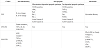
In sum, exercise training suppressed ovariectomy/diabetes-induced cardiac inflammatory status (decreases in Protein oxidation and TNF-α/ IL-10, increase in GSH/GSSG) as well as suppressed ovariectomy/hypertension-induced cardiac anti-oxidative stress (increases in SOD and Catalase) and inflammatory status (decreases in TNF-α, p-IKKα/ß, p-NFkB, COX-2). Exercise training suppressed ovariectomy-induced cardiac Fas dependent apoptotic pathways (decreases in t-Bid, Bad, Bax, Bak, Active Caspase 9, and Active Caspase 3) and mitochondria dependent apoptotic pathways (decreases in TNF-α, TNFR1, Fas, FADD, active Caspase 8, active Caspase 3) in ovariectomized rat models. Exercise training attenuated ovariectomy-induced cardiac fibrosis and fibrotic pathways (decreases in Collagen I, TGF-ß, p-Smad 2/3, CTGF, t-PA, MMP9) (Table 1). The findings might provide one possible mechanism of exercise training on preventing heart failure in menopausal or bilateral oophorectomied women through anti-inflammatory, anti-apoptotic, and anti-fibrotic pathways. Menopausal women should be highly aware of the progressive development in cardiac abnormality or heart failure, as well as they should devote themselves to exercise training and lifestyle modification.
Competing Interests
The authors declare that they have no competing interests.