


1. Introduction
Early prenatal and postnatal brain development represents one of the most critical and determinant of the human life. The neural effects of the first neurosensorial experiences established during the intrauterine and early postnatal period have been related to long-term developmental outcomes [1]. At birth, mother and child are actively involved during labour and delivery, when a synergic activation of physiological and biochemical mechanisms, both maternal and fetal, is necessary to ensure a normal perinatal outcome [2,3]. A growing interest has been paid to the factors that regulate or interfere with the intrauterine fetal well-being and the early weeks of life according to their role on short and long-term neuro developmental outcomes [4]. Herein we provided an overview of the main gestational and perinatal factors and/or events and their correlations with brain development. We also reviewed the main determinants of neurodevelopment and the influence of several environmental and biological factors occurring early in life.
2. Neurodevelopment is a Complex Fine-regulated Pathway
Neurodevelopment is a dynamic process highly regulated by the interaction between two main driving forces, nature and nurture [4]. Genes and environment mutually reinforce in developmental progression with the most prominent expression during ‘critical period’ or windows’ [1]. Hundreds of genes are involved into the main processes of human brain development from the early formation of neural tube up to the subsequent formation of the prosencephalon. Specific molecular pathways have been related to the forebrain development such as sonic hedgehog signalling pathway ( Shh) and a nodal pathway, respectively ventralizing and dorsalizing molecules, that, in their turn activate other genes and transcription factors ( TGIF, TDGF1, FAST1, Z1C2) and whose defects have been detected in patients with holoprosencephaly, for example genes involved into forebrain development and related to early [5]. Neural proliferation, migration, organization and myelination lead to the proper functioning of the intricate circuitry in the human brain and result from the complex interaction between environmental and genetic factors. Several genes were related to neuronal migration and, consequently, to the failure of this fine-regulated mechanism such RELN, LYS1, DCX, ARX, all detected in patients with lissencephaly [6]. You should mention about genetic factors and which genes play role on neurodevelopment. A critical period is a specific time-window associated to a highly expressed neural plasticity leading to easier and faster achievement of a given skill, e.g. walking or babbling. Neural networks in these specific periods are prone to be changed and shaped according to the environmental requests, leading to permanent learning [7]. Critical periods are time-limited and after their closure, the same experience no longer elicits the same degree of neural plasticity [7,8]. The time-onset of the critical period and its length are mainly influenced by the age of the child, however, they are highly related to the experience and environmental stimuli that sustain and prolong neural plasticity [7]. Environmental stimuli occurring early in development have been thought to induce brain structural changes such as in cortical width and weight, neuronal volume, dendrites arborization, axonal length and synaptic number and volume [1]. These evidences supported the need of identifying all factors early interfering with neurodevelopment and their related implications [9].
3. Gestational Factors
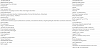
Several maternal and/or fetal factors have been reported to be specifically associated to the etiopathogenetic mechanisms underlying neonatal brain damage (Table 1). According to the type and time of occurrence we distinguish between:
- Antepartum and intrapartum demographic factors
- Environmental factors (“nurture”)
- Biomarkers
4. Antepartum and Intrapartum Factors
4.1 Antepartum factors
4.1.1 Clinical and demographic factors
Several maternal and fetal conditions may expose the unborn child to neonatal encephalopathy (NE) and subsequent cerebral palsy (CP). Maternal age, older than 40 and younger than 15 years, was associated to a higher rate of NE and CP. Diagnosis of epilepsy or other chronic neurological disorders (e.g. multiple sclerosis) have been associated to a double risk of NE [10]. Other factors include previous treatment for infertility, educational level, maternal type of work and marital status [11] (Table 1). Dietary habits of the mother such as vegetarian or vegan regimens might represent a prenatal risk of NE if not adequately balanced [12].
4.1.2 Obstetrical and gynecological diseases
Maternal genitourinary diseases may be associated to antepartum and intrapartum adversities. Bacterial vaginosis is the most common infection of the genitourinary tract of childbearing women, significantly associated to an increased risk of premature birth and perinatal transmitted infectious diseases [13]. It is characterized by an alteration of vaginal microbiota and an increase of pathogen agents such as Gardnerella vaginalis, Atopobium vaginae, Mobiluncus spp., Bacteroides spp. And Prevotella spp [14]. Tibaldiet reported that 77% of vaginal swabs in pregnancy resulted to be positive for specific agents [13].
Uterine structural anomalies such as septate or bicornute uterus, are associated to an increased risk of miscarriages and preterm birth [14]. Uterine benign tumors may be enlarged during pregnancy due to the hormonal stimulation thus increasing the risk of preterm birth if not treated before conception [15]. Structural and functional placental defects may be significantly associated to NE. Placenta previa is a condition in which the placenta implants in the poorly vascularized lower uterine segment that may results in inadequate uteroplacental perfusion and adverse neonatal outcome. This abnormal placentation may lead to severe peripartum hemorrhage and risk of maternal and fetal morbidity and mortality. Infectious placental diseases such as corioamnionitis, are also associated with preterm birth and NE [16] (Table 1).
4.1.3 Systemic diseases
Pre-eclampsia is an obstetrical emergency characterized by edema, proteinuria and hypertension whose etiopathogenetic mechanism seems to be immune-related and triggered by the fetus [17]. Acute clinical manifestations include severe hypertension, cerebral hemorrhage, intravascular disseminated coagulation, renal failure, and premature rupture of membranes [17]. Treatment of this severe, life-threatening syndrome is often associated with fetal delivery and pharmacological treatment. If pre-eclampsia occurs before the gestational term age, premature delivery will be performed [18].
Maternal chronic diseases such as diabetes, arterial hypertension, cardiopathies, autoimmune disorders, obesity have been reported to a 9-fold increased risk of NE, higher than pre-eclampsia (6-fold risk) [14]. Congenital TORCH infections (toxoplasma, rubella, cytomegalovirus, herpes, other) causing vertical transmission to fetus are an important risk factors of NE partially preventable with hygienic measures and, when available, immunization [19]. Smoking, alcohol and drug abuse are known maternal risk factors [20]. Congenital thrombophilic factors are associated to maternal thromboembolic complications with or without fetal involvement [21]. Prophylactic anticoagulant treatment (low molecular weight heparin) is commonly indicated to the at risk women [22] (Table 1).
4.1.4 Neurological and psychiatric diseases
The mother affected by epilepsy and/or other neurological and psychiatric disorders have an increased risk of adverse antepartum and intrapartum events [23]. Significantly reduced fetal growth has been evidenced in women with epilepsy when compared to those without epilepsy. The risk is also related to the antiepileptic treatment [24].
4.1.5 Fetal pathology
Fetal well-being is usually assessed according to international guidelines and represents a crucial tool forat-risk newborns early detection [25]. Several parameters need to be evaluated including fetal motility, fetal heart rate, placental and fetal flussimetria defects etc [26]. Cardiotocography allows the assessment of fetal heart activity as well as of fetal adaptation to uterine contractions. Tardive decelerations of the fetal heart rate may represent a red flag of uteroplacental insufficiency [27]. The presence of a congenital cardiomyopathy is a risk factor of cerebrovascular insult and neurodevelopmental impairment [28]. Multiple gestations are at risk of adverse maternal and fetal outcome, per se [29].
4.2 Intrapartum factors
Several intrapartum events are related to impaired neurodevelopment [29]. The most prominent factors include gestational age and neonatal birth weight. The risk is mainly increased in the lower epochs of gestational age, namely below 28 weeks, but also in late-term and post-term ages, after 42 weeks of gestational age, compared to the term age between 37 and 42 weeks [26,29]. Pregnancy is defined to be “prolonged” or Late Term after 41(+0) weeks, and post-term when it goes beyond 42(+0) weeks. The risk of maternal and fetal morbility has been reported to be progressively increased from 37(0-6) to 43(0- 6), although, a clear temporal cut-off of the risk has not been assessed [29]. As a whole, a newborn is considered at term when born between 37(+0) and 41(+6) weeks of gestation. Close antepartum monitoring and labour induction are the most used strategies to reduce perinatal morbidity and mortality due to late or post-term birth. The risk concerns both the fetus (intrauterine fetal loss, macrosomia, meconium aspiration syndrome etc) and the mother (emergency Caesarean section, perineal lesions, post-partum hemorrhage etc) [25]. The signs of fetal distress include meconium stained amniotic fluid, which causes aspiration pneumonia and respiratory distress, and also fetal bradycardia, altered gases in umbilical arterial blood, and other specific features evidenced by cardiotocography and ultrasound scans [30]. The onset of fever during labour is often associated to corioamnionitis, fetal tachycardia and neonatal sepsis and may represent a risk of NE. Premature rupture of membranes (PROM), characterized by the sudden rupture of the amniotic sac, bleeding and labour onset was related to an increased risk of NE [31]. Some conditions appear to facilitate the occurrence of PROM such as uterine and vaginal infections. Other predisposing factors include cervix abnormal lenght and patency, multiple gestations or traumas [31]. Maternal seizures occurring during labour and/or delivery may increase the risk of neonatal asphyxia [23]. Previous pregnancies especially with operative delivery increase the risk of maternal peripartum bleeding and uterine rupture [30]. Apgar score at 5 and 10 minutes has been related to risk of CP and epilepsy [32]. Mercuri et al. (2002) showed that 28% of newborn with NE and severe Apgar score (<3), presented with normal brain MRI scan, or showed minor white matter abnormalities, conversely, 95% of newborns with Apgar score <7 showed abnormal neuroimaging at 48hours of life [33]. Among 988 children diagnosed with CP at 5 years of age by Lie et al. (2010), 11% have had Apgar score <3, but only 0.1% received an Apgar score equal to 10 with a 53-fold increased risk in the lower Apgar group [34]. Other intrapartum risk factors include: neonatal sepsis, bronchopulmonary dysplasia, necrotizing enterocolitis (stage II/III), neonatal hypoglycemia and hyperbilirubinemia [30]. Neonatal seizures are an independent risk factor associated to NE and CP [35].
Several antepartum and intrapartum risk factors may occur either isolated or in association. Some authors have reported a growing grading of risk according to the association of two or more risk factors compared to the occurrence of an isolated factor. The time-onset of brain damage, namely if it occurs antepartum, intrapartum or both, is still debated. NE is diagnosed when neurological symptoms onset is within 24 hours after birth and after an acute insult leading to hypoxic-ischemic brain damage [36]. However, brain lesions are not necessarily related to perinatal insults [33]. Antepartum factors may predispose to subsequent intrapartum adversities conversely, the latter cannot be enough to cause brain damage and neurodevelopmental sequelae. Cowan et al. reported that hypoxic-ischemic encephalopathy (HIE) takes its origin before birth, but an intrapartum secondhit is necessary to establish the damage35. Maternal age, previous number of pregnancies and obstetrical complications, low or high estimated neonatal birth weight may be taken into account to plan a close monitoring during pregnancy, labour and delivery. A retrospective study (Badawi et al. (1998), showed the absence of any intrapartum adversities in 70% of a population of children with NE, thus, antepartum period appeared to be determinant in NE etiopathogenesis [29].
5. Early Post-natal Experiences and Developmental Care
A growing literature has been focused on the effects of the early experiences on neurodevelopment in newborns, especially those mainly exposed to at-risk conditions such as preterm babies. Even in the absence of perinatal adversities, preterm newborns are potentially exposed to visual, auditory, tactile and painful early atypical experiences that may represent a neurodevelopmental risk themselves [4,37]. The Newborn Individualized Developmental Care and Assessment Program (NIDCAP) has been conceived on these grounds in 1980 [38] with the aim to promote the ‘alchemy’ between mother and newborn by favouring reciprocal dyadic interactions as well as handling time. NIDCAP is an individualized approach for holding and caring, based on neonatal behavioral observations and planning a personalized care program (www.nidcap.org) [38]. The treatment starts when the newborn reaches the NICU and will continue throughout the hospitalisation and after discharge as a home-therapy. The Kangaroo Mother Care (KMC) centralizes the role of the family involving every member in the care of the newborn and emphasizing the baby-parents physical and emotional relationship. This supportive care model has been created to reduce the impact of the transition between the intrauterine prenatal environment and the NICU conditions, to promote the skin-to-skin contact and to strengthen the mother-baby diad [39]. Another important approach to improve neonatal care is the massage. Randomized controlled trials showed that the newborns receiving massage therapy have a significant weight gain compared to those who do not receive it [40]. Massage therapy for 5-10 days has been shown to increase newborn’s weight by 21-48% and decrease hospitalisation length between 3 to 6 days compared to controls [41-43]. On the other hand, reduced rates of anxiety and depression disorders have been reported when the mothers practise the massage for themselves [45]. Moreover, the use of polyunsaturated oils to perform the massage has been related to the weight gain [45]. Other beneficial effects following a massage with moderate pressure encompassed the improvement of gastrointestinal motility, the insulin and IGF-1 levels [46].
6. Biomarkers
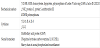
Several biomarkers possibly associated to neonatal brain hypoxicischemic damage have been investigated and related neuroprotection strategies have been proposed. By definition, a biomarker should be rapid and easy to use, as well as able to predict both short and long term outcome. However, the majority of biomarkers are not specifically related to HIE and poorly predictive of the neurological outcome [47]. Moreover, the optimal time of measurement and its relevance in clinical practice are poorly defined as yet. Serum, urine and cephalo spinal fluid (CSF) biomarkers have been assessed in newborns with HIE, however they showed little specificity for neural tissues (Table 2).
6.1 Biochemical markers
Nagdyman et al. showed the increase of creatine kinase brain isoform (CK-BB) and S100b but not the neuron-specific enolase (NSE) in term newborn with HIE, although none of them was significantly related to morbidity and mortality at 20 months of age [48]. In contrast, Gazzolo et al. showed that urine S100b is related with severe outcome at 12 months of age [49]. Celtik et al. reported that serum increase of NSE is not related to neurological outcome [50], whereas Walsh et al. evidenced that serum and cordonal CK-BB levels measured at 6 hours of life are associated with seizures’ onset and neurological abnormalities at 7 months of age in 77% of newborns with HIE [51]. Serum NSE and S100b during the first 72 hours after birth have been described in newborn with HIE underlying hypodermic treatment and resulted to be highly predictive of brain lesions at MRI scans at 14 days of life, as well as mortality [52]. A direct correlation between plasma homocysteine levels and methylenetetrahydrofolate reductase (MTHFR) polymorphisms (A1298C, C677T) has been reported in preterm and term newborn with NE due to HIE [53]. Homocysteine is a sulphur amino acid involved in methionine metabolism. Cofactor deficiency or genetic polymorphisms of the catabolic enzyme MTHFR will contribute to the increase of homocysteine plasma levels and increase oxidative stress and excitoxicity [53,21]. Increased homocysteine plasma levels and MTHFR polymorphisms have been associated to specific white matter abnormalities in both preterm and term infants [21]. Deficiency of other coagulative factors such as protein C, protein S and antithrombin III, elevated plasma lipoproteins and polymorphisms of Leiden factor V G1691A and factor II (prothrombin) G20210A, have been associated to cerebral venous thrombosis in the newborn [21].
6.2 Immuno-inflammatory markers
Several immuno-inflammatory markers have been reported in newborns with HIE. Bartha et al. showed that the serum levels of IL-1b, IL-6, IL-8, and TNF-a, were related to increased lactate and choline in deep nuclei in brain magnetic resonance spectroscopy, thus suggesting abnormal oxidative metabolism. However, this finding was not confirmed by brain MRI scan at 6 days of life [54]. In the same study, children with neurodevelopmental disorders at 30 months had neonatal increased levels of IL-1b, IL-6, IL-8, and lower levels of IL- 12, and no changes in IL-9, IL-13, and TNF levels [54]. Conversely, Savman et al. failed to show correlations between IL-8 levels and neurodevelopmental outcome [55]. No correlations arose between cytokines’ levels and severity of HIE neither did any difference emerge between hypotermic treatment and rewarming [56]. In contrast, Roka et al. showed that hypothermia was associated to reduction of IL-6 and IL-4 [55], and also an adverse neurodevelopmental predictive role after hypothermia has been related to the levels of pro-inflammatory cytokines IL-1, IL-6, e IL-8 [56].
6.3 Specific cerebral biomarkers
Some novel specific cerebral biomarkers have been identified. glial fibrillary acid protein (GFAP) is an intermediate filament in astrocytes, ubiquitin carboxyl-terminal hydrolase L1 (UCHL1) is a cytoplasmic neuron-specific expressed in dendrites, and the phosphorylated axonal neurofilament is the main cytoskeleton axonal and dendritic protein. These cerebral biomarkers have been related to traumatic brain injuries and to the severity of the cerebral damage [57]. Cordonal GFAP and UCHL1 appeared to be related to the severity of HIE, however, only GFAP resulted to be significantly increased compared to UCHL1 in serial assessments at 6-24, 48, 72 and 78 hours of life [56].
7. Conclusions
Several pre-natal and post-natal factors may interfere with neurodevelopment and have been related to early brain damage. Both genetics and experience play a crucial role on brain structural and functional development, and, specific prenatal and early postnatal care models have been defined. An increased focus on the at-risk pregnancies is a very crucial point to implement measures of prenatal prevention for brain damage and to plan early follow-up programs and interventions in newborns.
Competing Interests
The authors declare that they have no competing interests.