


1. Introduction
Vitamin D is a secosteroid associated with peripheral calcium homeostasis and nervous system function [1]. Significant changes in maternal vitamin D and calcium metabolism occur during pregnancy to provide calcium for fetal bone mineral accretion [2]. It is important that 25OHD readily crosses the placental tissue to the fetus [3,4]. Low 25OHD levels in the maternal and cord blood have been found to be significantly associated with decreased birth weight of infants [5]. Vitamin D deficiency is common in preterm infants [6] and vitamin D supplementation in women at high risk of vitamin D deficiency leads to improved neonatal handling of Ca [2]. Many studies have shown that maternal vitamin D level might affect fetal growth.
Autism spectrum disorders are developmental disorders associated with a high individual and social burden, but their aetiology is poorly understood. We previously reported that vitamin D supplementation for 9 months might ameliorate typical clinical symptoms in children with autism spectrum disorder [7]. Vitamin D may have an important role in the development of the brain.
In comparison with serum 25OHD concentration in non-pregnant women or women with uncomplicated pregnancies, half of urinary 25OHD was excreted in uncomplicated pregnant women at 15 weeks’ gestation [8]. Given this, serum vitamin D state was quantified from maternal urinary vitamin D.
PC12 cells represent a useful in vitro model of neuronal differentiation with various neurobiological properties [9]. Exposure of nerve growth factor (NGF) cause PC12 cells to differentiate into sympathetic neuron-like cells that exhibit increased neurite outgrowth and synapses. However, to our knowledge there has been no study analyzing the effects of vitamin D on nerve growth in vitro. Another aim of the present study was therefore to identifywhether25OHD is associated with NGF-induced nerve growth.
2. Materials and Methods
2.1 Subjects and Setting
Prior to this study, approval was obtained from the ethics committee of Doshisha Women’s College of Liberal Arts (project registration number in 2018: 2018-26). We enrolled 78 pregnant women with planned deliveries from March to August in 2019. Maternal urine samples were obtained and body weight, height, age and gravidity in maternal and child health screening in the clinic between 11th February 2019 and 15th March 2019 were recorded. Urine specimens were classified into two groups in consideration of the number of days from the urine collection day to the delivery day, and stored at -80°C before analysis. One group was more than 90 days to delivery (the late first and second trimester) and the other was within 90 days to delivery (the third trimester).
2.2 Serum 25OHD and creatinine
Urinary 25OHD and creatinine were analyzed using a commercially available ELISA kit (Immundiagnostik AG, Germany) and colorimetric reaction kit using Jaffe reagent (QuantiChrom TM, CA, USA), respectively. The 25OHD concentrations were normalized for urinary creatinine (ng/mg creatinine).
2.3 Newborn anthropometry
Newborn anthropometry (height (cm), head circumference (cm), chest circumference (cm) and birth weight (kg)) of babies delivered naturally were measured by skilled midwives.
2.4 Cell culture and assay
PC12 cells obtained from the Riken Cell Bank (Ibaraki, Japan) were maintained in DMEM supplemented with 10% fetal bovine and horse serum (Gibco, Life Technologies) and 1% penicillin-streptomycin (Sigma). The cells were then incubated at 37°C in an atmosphere of 5% CO2/95% air. PC12 cells were seeded at a density of 1.5 x 10⁵ cells [9]. Following 24 h of incubation, PC12 cells were treated with 0 ng/mL NGF with or without 6 nmol/L of 25OHD. The ACE activity was measured for an index of the differentiation of the PC12 cells and expressed as absorbance mIU of mg protein. AchE activity and protein were analyzed using a commercially available kit (AmpliteTM Colorimetric kit, AAT Bioquest, USA) and PierceTM BCA protein assay kit (Thermofisher Scientific, USA), respectively.
2.5 Statistical analysis
The differences between urinary 25OHD and newborn anthropometry were evaluated using the t-test. AChE activity was expressed as mean ±SD and evaluated using the t-test. A p-value of < 0.05 was considered to be statistically significant. Analyses were carried out using SPSS 21 for Windows (IBM, Japan).
3. Results and Discussion
3.1 Study subjects
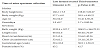
The characteristics of the study subjects are shown in Table 1. There were no significant differences in delivery characteristics between the two groups classified by the times of urine specimen collection. The height and the weight at the time of the birth were a little bigger than the mean values shown by the Ministry of Health, Labor and Welfare. These data showed that the deliveries in this study were typical Japanese deliveries.
3.2 Newborn anthropometry outcomes and urinary 25OHD concentration in pregnancy
The associations between newborn length, chest circumference and birth weight and the urinary 25OHD concentration of mothers in the late first and second trimester and the third trimester were not significant. A slightly positive association between urinary 25OHD and birth head circumference was observed in the late first and second trimester (r= 0.358), but not in the third trimester (Figure 1).
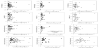
The vitamin D receptor is found in multiple brain regions of the neonatal central nervous system [10,11]. Maternal vitamin D supplementation from the 2nd half of pregnancy until birth does not influence newborn growth [12]. Our study showed a slightly positive association between birth head circumference and urinary 25OHD concentration for the late first and second trimester. These results suggest that vitamin D levels in early pregnancy (1st trimester) are associated with newborn head size.
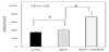
3.3 Enhancement of NGF-induced neuronal differentiation in PC12 cells by 25OHD
Six nmol/L of 25 OHD (=1/5 of serum level) enhanced nerve growth factor induced differentiation in PC12 cells (Figure 2).
Vitamin D plays a role in brain developmental mechanisms [13]. Lower maternal serum 25 OHD in the first trimester is associated with a higher autism risk [14]. These results suggest that preventing vitamin D deficiency in early pregnancy is essential to decrease the risk of autism. Vitamin D deficiency is highly prevalent in the winter months, seen in 64% of pregnant women [15]. Instruction with advice on how to supplement vitamin D by sunbathing and vitamin D-rich food is necessary.
This is a preliminary study with a very small number of subjects. Further study with larger numbers of subjects is warranted, and could reveal optimal 25 OHD levels for preventing vitamin D deficiency in the late first trimester.
4. Conclusion
These findings show that advice on vitamin D supplementation in the late first and second trimester is beneficial for the healthy development of the fetal head circumference and neural system.
Competing Interests
The authors declare that they have no competing interests.
Author Contributions
Dr. Hasegawa was responsible for the study conception, design,
analysis, interpretation of data, and drafting of the manuscript.
Dr. Manabe was responsible for the data acquisition and proof reading
of the manuscript.
Dr. Izumi was responsible for the data acquisition and proof reading
of the manuscript.
Ms.Mochizuki was responsible for proof reading of the manuscript,
and participated in the data analysis.
Acknowledgments
The authors acknowledge Dr.Seiji Fujita of Fujita Ladies Clinic (Kyoto, Japan) for his kind help.