Profile
International Journal of Laboratory Medicine & Research Volume 1 (2015), Article ID 1:IJLMR-107, 3 pages
http://dx.doi.org/10.15344/2455-4006/2015/107
Commentary
Apolipoprotein E-knockout Mice as a Lifestyle-related Disease Model of Atherosclerosis and Non-alcoholic Fatty Liver Disease

Masa-Aki Shibata1*, E. Shibata2, S. Fujioka1 and M. Harada-Shiba2

1Laboratory of Anatomy & Histopathology, Graduate School of Heath Sciences, Osaka Health Science University, Japan
2Department of Molecular Innovation in Lipidology, National Cerebral & Cardiovascular Center Research Institute, Japan
Prof. Masa-Aki Shibata, Laboratory of Anatomy & Histopathology Graduate School of Heath Sciences, Osaka Health Science University, 1-9-27, Temma, Kita-ku, Osaka, 530-0043, Japan, Tel: +81-6-7506- 9046; Fax: 81-6-6352-5995; E-mail: masaaki.shibata@ohsu.ac.jp
12 October 2015; 14 October 2015; 16 October 2015
Shibata MA, Shibata E, Fujioka S, Harada-Shiba M (2015) Apolipoprotein E-knockout Mice as a Lifestyle-related Disease Model of Atherosclerosis and Non-alcoholic Fatty Liver Disease. Int J Lab Med Res 1: 107. doi: http://dx.doi.org/10.15344/2455-4006/2015/107
This work was supported by Grants-in-Aid for Scientific Research from the Japanese Ministry of Health, Labor and Welfare (H23- seisakutansaku-ippan-004. A member of this research: Dr. M.A. Shibata. The research representative: Dr. M. Harada-Shiba).

Hypertension, hyperlipidemia, diabetes, and obesity are the "deadly quartet" of lifestyle related diseases, those whose onset and slow progression are the result of choices made in everyday living. Each condition is bad enough alone, but combined they significantly increase the risk of death. Atherosclerotic lesions are a frequent hallmark of lifestyle-related disease; they develop silently and often go undetected until their condition and location precipitate myocardial and/or cerebral infarction and, not infrequently, sudden death.

Atherosclerosis and its complications are among the leading causes of mortality in Western countries [1] and a similar trend is reported in Japan [2]. Non-alcoholic fatty liver disease (NAFLD) is the most common liver disease in both adult and pediatric populations [3,4]. Clinically it is divided into simple fatty liver without inflammation and fibrosis and steatohepatitis; if left untreated, however, progression to steatohepatitis is nearly certain. Severe NAFLD due to diabetes mellitus and obesity commonly advances to inflammatory non-alcoholic steatohepatitis (NASH), a precursor of cirrhosis and ultimately hepatocellular carcinoma [6]. Since NAFLD and atherosclerosis share common molecular mediators, NAFLD itself might play a crucial role in the development and progression of atherosclerosis [5].

Animal studies are crucial to the development of therapeutic drugs for atherosclerosis and NAFLD. A gene-targeted mouse model of atherosclerosis has been created at the Jackson Laboratory in Bar Harbor, Maine, USA, in which C57BL/6J mice homozygous for the ApoEtm1Unc mutation demonstrate a functional knockout of the antiatherogenic apolipoprotein E (ApoE) gene involved in cholesterol metabolism [7,8]. This ApoE-knockout (ApoE-KO) mouse has been widely used in atherosclerosis research because of its propensity to spontaneously develop hypercholesterolemia and atherosclerotic lesions similar to those found in humans [9,10]. In addition to atherosclerotic lesions, ApoE-KO mice over 6-months of age develop hepatic steatosis and fibrosis [11].

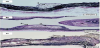
Atherosclerotic lesions in ApoE-KO mice are histopathologically classified into 3 categories by stages [12]: i) early, ii) progressive/ advanced, and iii) combined lesions. Early lesions are simple accumulations of foamy cells in aortic endothelial epithelium (Figure 1A). Progressive lesions are composed of foamy cell accumulations, fibrous caps and lipid cores (Figure 1B). Combined lesions show accompanying calcification and/or ossification (Figure 1C), with occlusion of the lumen in progressive lesions.

figure 1
Figure 1: Histopathology of atherosclerotic lesions in male ApoE-KO mice fed normal diet (A,B) or high fat diet containing 1.25% cholesterol (C). A) Early lesion at 20-weeks of age. Note appearance of foamy cells (1) on endothelial wall. B) Progressive lesion in a 38-week old mouse with foamy cells (1), fibrous cap (2) and lipid core (3). C) A combined lesion in a 23-week old mouse showing ossification and chondrofication (4), demarcated by the broken line. [Elastica van Gieson stain for elastic fiber. A) Scale bar=50 μm; B,C) Scale bar=100 μm].

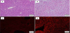
Histopathological findings in ApoE-KO livers are presented in Figure 2. Compared to normal liver cells in wild-type mice (Figure 2A), variable hepatocyte size with cytoplasmic vacuolization are evident without the presence of inflammatory cells and fibrosis (Figure 2B). Frozen liver sections stained with Nile Red for demonstration of lipids showed that, while normal liver shows almost no signals (Figure 2C), ApoE-KO livers demonstrate numerous lipid droplets within hepatocytes (Figure 2D), indicating fatty metamorphosis similar to simple fatty liver in human NAFLD.

figure 2
Figure 2: Histopathology of liver in ApoE-KO male mice fed normal diet. A) Normal liver in a 20-week old mouse; note the uniform hepatocyte size with little cytoplasmic vacuolization. B) Fatty liver in a 24-week old animal showing variable hepatocellular size, cytoplasmic vacuolization and lipid droplets; note the lack of inflammatory cells and fibrosis. C) Frozen section of the normal liver of a 20-week old mouse, stained with Nile Red. Very few intracellular lipid droplets are seen. D) Frozen liver section from a 24- week old male with fatty metamorphosis, equivalent to simple fatty liver in human NAFLD. Note the abundance of intracellular lipid accumulation shown with Nile Red staining. [A,B - H&E; C,D- Nile Red fluorescent stain (Wako Pure Chemical Industries, Inc., Osaka, Japan; excitation wavelength 485-530 nm, fluorescence wavelength 525-605 nm). A-D Scale bar=50 μm].

In a practical sense, mice are the preferred research animal in terms of economical space and husbandry requirements, and the ability to generate specific gene equivalency between mouse and man is extremely useful in translational research. Because both atherosclerosis and NAFLD normally occur in this line and are promoted by dietary fat, ApoE-KO mice provide a functional model to aid in the elucidation of molecular mechanisms and the development of therapeutic agents for treatment of human atherosclerosis and NAFLD.

Competing Interests

The authors have no competing interests with the work presented in this manuscript.


References

  1. World Health Organization. The top 10 causes of death in the world, 2000 and 2012. Fact sheet No 310; updated May 2014. View
  2. Ministry of Health, Labor and Welfare in Japan Annual Report 2012. The top 10 causes of death in Japan , 2012. View
  3. Review Team: LaBrecque DR, Abbas Z, Anania F, Ferenci P, et al. (2014) World Gastroenterology Organisation global guidelines: Nonalcoholic fatty liver disease and nonalcoholic steatohepatitis. J Clin Gastroenterol 48: 467-473. View
  4. Lavine JE, Schwimmer JB (2004) Nonalcoholic fatty liver disease in the pediatric population. Clin Liver Dis 8: 549-558, viii-ix. View
  5. Targher G (2007) Non-alcoholic fatty liver disease, the metabolic syndrome and the risk of cardiovascular disease: the plot thickens. Diabet Med 24: 1-6. View
  6. Cohen JC, Horton JD, Hobbs HH (2011) Human fatty liver disease: old questions and new insights. Science 332: 1519-1523. View
  7. Piedrahita JA, Zhang SH, Hagaman JR, Oliver PM, Maeda N (1992) Generation of mice carrying a mutant apolipoprotein E gene inactivated by gene targeting in embryonic stem cells. Proc Natl Acad Sci U S A 89: 4471-4475. View
  8. Plump AS, Smith JD, Hayek T, Aalto-Setälä K, Walsh A, et al. (1992) Severe hypercholesterolemia and atherosclerosis in apolipoprotein E-deficient mice created by homologous recombination in ES cells. Cell 71: 343-353. View
  9. Coleman R, Hayek T, Keidar S, Aviram M (2006) A mouse model for human atherosclerosis: long-term histopathological study of lesion development in the aortic arch of apolipoprotein E-deficient (E0) mice. Acta Histochem 108: 415-424. View
  10. Meyrelles SS, Peotta VA, Pereira TM, Vasquez EC (2011) Endothelial dysfunction in the apolipoprotein E-deficient mouse: insights into the influence of diet, gender and aging. Lipids Health Dis 10: 211. View
  11. Stachowicz A, Olszanecki R, Suski M, Wiśniewska A, Totoń-Żurańska J, et al. (2014) Mitochondrial aldehyde dehydrogenase activation by Alda-1 inhibits atherosclerosis and attenuates hepatic steatosis in apolipoprotein E-knockout mice. J Am Heart Assoc 3: e001329. View
  12. Shibata MA, Shibata E, Fujioka S, Harada-Shiba M (2015) Atherosclerosis in apolipoprotein E-knockout mice as a model of human disease. Austin J Cardiovasc Dis Atherosclerosis 2: 1011.