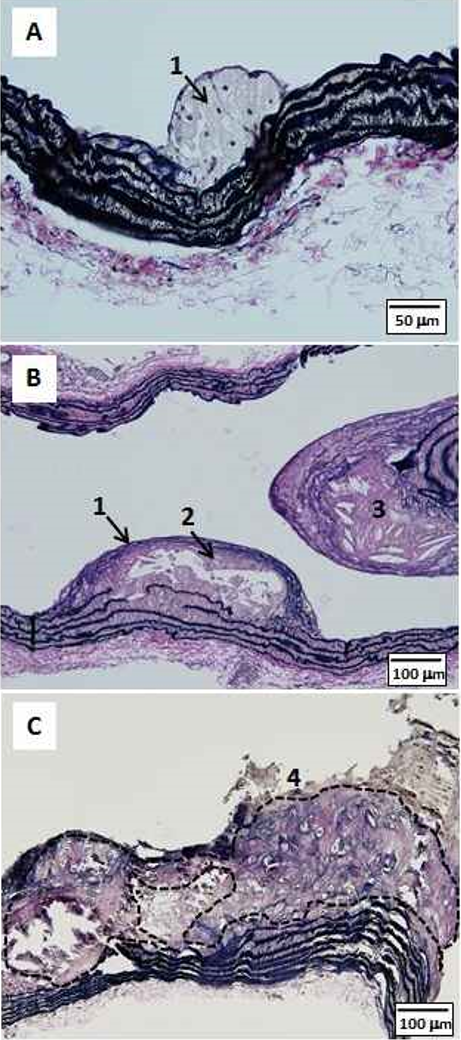
Figure 1: Histopathology of atherosclerotic lesions in male ApoE-KO mice fed normal diet (A,B) or high fat diet containing 1.25% cholesterol (C). A) Early lesion at 20-weeks of age. Note appearance of foamy cells (1) on endothelial wall. B) Progressive lesion in a 38-week old mouse with foamy cells (1), fibrous cap (2) and lipid core (3). C) A combined lesion in a 23-week old mouse showing ossification and chondrofication (4), demarcated by the broken line. [Elastica van Gieson stain for elastic fiber. A) Scale bar=50 μm; B,C) Scale bar=100 μm].