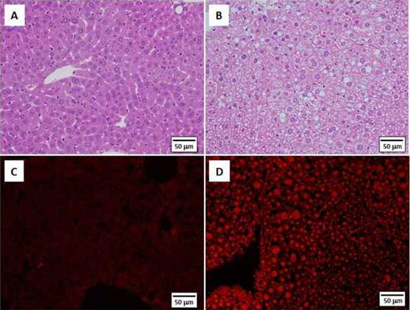
Figure 2: Histopathology of liver in ApoE-KO male mice fed normal diet. A) Normal liver in a 20-week old mouse; note the uniform hepatocyte size with little cytoplasmic vacuolization. B) Fatty liver in a 24-week old animal showing variable hepatocellular size, cytoplasmic vacuolization and lipid droplets; note the lack of inflammatory cells and fibrosis. C) Frozen section of the normal liver of a 20-week old mouse, stained with Nile Red. Very few intracellular lipid droplets are seen. D) Frozen liver section from a 24- week old male with fatty metamorphosis, equivalent to simple fatty liver in human NAFLD. Note the abundance of intracellular lipid accumulation shown with Nile Red staining. [A,B - H&E; C,D- Nile Red fluorescent stain (Wako Pure Chemical Industries, Inc., Osaka, Japan; excitation wavelength 485-530 nm, fluorescence wavelength 525-605 nm). A-D Scale bar=50 μm].