


1. Introduction
Cardiovascular autonomic failure (CAF) is a frequent non-motor symptom of Parkinson’s disease (PD), occurring in up to 30% of cases [1]. The key feature of CAF is orthostatic hypotension (OH), defined as the decrease of systolic blood pressure (BP) ≥ 20 mmHg or diastolic BP ≥ 10mHg upon standing or 60° head-up tilting [2]. This is frequently accompanied by supine and/or nocturnal hypertension, as the result of global alteration of BP control[3,4] .
Several research groups suggested the association between BP dysfunctions and cerebrovascular damage in PD [5]. Thus, CAF was coupled with a higher burden of cerebral white matter lesions, i.e. regions of altered parenchymal signal at CT/MRI scan due to chronic cerebrovascular damage. Notably, white matter lesions contribute to neurological disability in PD, in particular by aggravating symptoms scarcely responsive to dopamine replacement therapy such as postural instability and cognitive impairment [6].
Despite the potential relevance of these previous findings, several clues need to be elucidated. Indeed, results in the literature suffer from biases due to the great variability of duration and severity of BP deregulation in PD. Moreover, the pathophysiological mechanisms underlying the association between BP fluctuations and cerebrovascular damage in PD are still undefined. Eventually, very few data are available on cerebral vasoreactivity (CVR) in PD patients [7,8].
CVR refers to the dilatory response of cerebral arterioles to elevation of CO2 partial pressure [9]. Reduced arteriolar tone leads to increased blood flow velocity in cerebral vessels aimed at “washing out” CO2 from the brain, thus maintaining pH and cerebral homeostasis. CVR is a chemically-based process initiated within the endothelium, and is viewed as a marker of preserved endothelial functions [10]. CVR may be reduced in individuals suffering from hypertension [10]. Moreover, reduced CVR is an independent predictor for stroke in the setting of severe carotid artery stenosis [11].
In outpatient subjects, hypercapnia may be obtained by holding breath, and the subsequent variations in blood flow velocities can be monitored by transcranial doppler sonography of the main cerebral vessels. The percentage increase of blood flow velocity divided by the duration of apnoea, defined as “breath holding index” (BHI), has been widely utilized to estimate CVR (see also “Methods” sections) [9].
To gain further knowledge on the influence of autonomic disturbances on cerebral hemodynamics in PD and to evaluate BHI as a possible marker in this context, here we measured BHI in PD patients with and without CAF as compared to healthy subjects.
2. Materials and Methods
Twelve subjects diagnosed with PD according to international criteria [12] and 11 healthy subjects were enrolled. All subjects underwent clinical evaluation to exclude major metabolic (e.g., diabetes, dyslipidemia), cardiovascular or neurological co-morbidity, neuroimaging study (CT or MRI) to exclude major strokes, and duplex sonography to exclude significant carotid artery stenosis (>30%). The study protocol was approved by the “Santa Lucia Foundation” IRCCS Ethical Committee and conducted in accordance with the Declaration of Helsinki. Each participant signed informed consent prior to enrollment.
To minimize drug influence, we performed all evaluations before the first morning dose of dopaminergic drugs and/or anti-hypertensive medication. On the examination day subjects were also invited not to drink any beverage containing caffeine, theine or taurine.
To evaluate CVR, hypercapnia was reproduced by holding breath. The subsequent increase in mean blood flow velocity is expressed as a percentage and divided by the seconds of apnoea. The obtained parameter is known as “breath holding index” (BHI) (see below) [8].
Variation in blood flow velocities were monitored with transcranial doppler ultrasound. Middle cerebral arteries were bilaterally insonated through the temporal bone window using a headband, and blood flow velocities were continuously recorded after 10 minutes resting in supine position before performing the breath holding. Afterwards, a standing test was performed for 3 min, and mean blood flow velocities were likewise recorded prior and during a further breath holding test.
The presence of OH in PD patients was evaluated by means of the standing test according to the current international diagnostic criteria [2].
The statistical analysis was performed using SPSS, version 20.0. Normal distribution of data was checked using the Shapiro-Wilk test. Quantitative variables were analyzed by means of unpaired t-test if they showed a normal distribution or Mann-Whitney-U test if not. Fisher’s exact test was used to compare categorical variables. P values <0.05 were considered statistically significant.
3. Results
3.1 Comparison between PD patients and controls
The demographic and clinical data of PD patients and controls are shown in Table 1. The prevalence of hypertension or smoking habit did not differ between PD patients and controls. PD patients displayed significantly higher BP and heart rate values together with a trend towards lower basal blood flow velocity values. At CVR assessment, BHI was significantly lower in the PD patients with respect to healthy subjects.
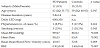
*statistically significant comparison.
3.2 Patients intra-group comparison
During the standing test, OH was diagnosed in 4/12 PD patients. These patients (OH+) had comparable disease duration, but they were older and received significantly lower daily amount of dopamine replacement therapy as compared to OH- (see table 2). OH+ patients displayed a tendency towards higher supine mean BP values and significantly lower supine basal blood flow velocities. Nevertheless, in the CVR assessment both groups had similar BHI values. Upon standing, apart from the expected significant differences in BP values, heart rate and blood flow velocity variation did not display significant differences between OH+ and OH- patients (Table 2, “Difference standing-supine” section). OH+ patients displayed reduced mean blood flow velocity also upon standing comparing to OH-.
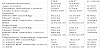
*statistically significant comparison.
4. Discussion
The results of this pilot study demonstrate significant difference of CVR between PD patients and controls. These findings are unlikely related to CAF, as OH+ and OH- PD patients displayed similar performances.
We applied BHI to challenge CVR because of its feasibility in outpatient setting. Furthermore, previous reports showed that such method is equivalent to other standardized tests, such as re-breathing techniques or acetazolamide challenge [9].
To our best knowledge, only one study dealt with BHI evaluation in PD patients previously [7]. Our OH+ and OH- PD patients displayed similar BHI values as compared to those by Camargo et al. [7]. However, our OH+ cases had significantly lower basal mean blood flow velocity with respect to OH- (Table 2). Furthermore, Zamani et al. [8] reported a blunted increase in blood flow velocities upon inhalation of CO2-enriched air in 34% of their PD patients, that was not related to the presence of OH. In that study, however, comparison with control subjects was not performed.
We acknowledge that the small sample size limits the power of our study. However, we believe that the present findings are strengthened by comprehensive pre-test evaluation to exclude the most relevant confounding factors (metabolic disorders, carotid artery stenosis, other causes of cerebrovascular disorders that may influence CVR). Furthermore, differently from previous reports [7], in our study OH was identified according to the current international guidelines after 3 min standing [2].
CVR is an endothelial-dependent mechanism that may be compromised in several disorders, such as hypertension, carotid artery stenosis and dementia[10,11] . The results of this pilot study, together with previous reports[7,8] , show altered CVR in PD patients, independently from the presence of CAF. Further, we provide preliminary evidence of lower basal blood flow velocity in OH+ as compared to OH- PD patients while supine and upon standing. These findings, therefore, show reduced CVR compliance in PD patients, that might play a role in the development of chronic cerebrovascular damage facilitated by the presence of cardiovascular autonomic failure. The pathophysiological basis and the prognostic relevance of these preliminary findings, however, remain to be established by future prospective studies on larger cohorts.
Competing Interests
The authors have no competing interests with the work presented in this manuscript.
Acknowledgments
This was an academic project; no external financial support was assigned.