


1. Introduction
Nuclear medicine technology provides both functional, which is aimed to quantify the physiological process, and anatomical information. Accordingly, in the field of the diagnostic imaging, importance of this technology has been increased [1-2].
In the nuclear medicine imaging two major kinds of devices have been used: (1) gamma camera or single photon emission tomography (SPECT) with collimator and (2) positron emission tomography (PET) with non-collimator. The monoenergetic gamma ray source such as most commonly used 99mTc (140 keV energy peak and half-life of 6 hours) is generally used in the gamma camera or SPECT [3-4]. For detecting gamma ray source, we should be used radiation detector. In the field of the nuclear medicine, NaI(Tl) scintillation detector and conventional or newly developed semiconductor detectors are frequently used. Among these detectors, NaI(Tl) scintillation and conventional semiconductor detector using silicon (Si) or germanium (Ge) have low intrinsic resolution and low detection efficiency problems, respectively. Especially, Si or Gehas high cost and is required additionally cooling system. To overcome these problems, newly developed semiconductor detectors based on cadmium zinc telluride (CZT) and cadmium telluride (CdTe) has been developed during recent years. These detectors can be detected gamma ray in room temperature condition without cooling system, unlike conventional semiconductor detector. Especially, the main advantages of CZT and CdTe semiconductor detectors were good energy resolution of approximately 3-6% at 140 keV and high sensitivity due to high detection efficiency. Also,the excellent spatial resolution can be acquired by using these detectors because intrinsic resolution is same to the pixel size [5-10]. In this regard, by offering better sensitivity and spatial resolution, gamma cameras using CZT and CdTe semiconductor detectors can overcome inherent limitations in image performance of the conventional gamma camera using Si or Ge semiconductor and NaI(TI) scintillation detectors.
In the gamma camera or SPECT system, collimators play two main roles in nuclear medicine imaging. The one, this imaging is formed by selective absorption of the emitted gamma rays in the body. Thus, it is essential to know gamma rays direction finder. The other, gamma camera systems can cut-off unnecessary gamma rays such as scattering and conduct selective absorption by using a collimator. For these reasons, collimators are one of the most important components of gamma camera systems. Also, because of the image performance such as sensitivity and spatial resolution that characterize collimators, collimator design markedly influences the image performance of the gamma camera system [11-14]. Among collimators, pinhole collimator with geometric magnification effect can be used for the pre-clinical gamma camera system to achieve high spatial resolution. Especially, in CZT or CdTe semiconductor detector system, the magnification effect has influence on intrinsic resolution. Thus, pinhole collimator has been used for very small objects such as the thyroid. In addition, sensitivity of pinhole system is improved when closing the distance between the pinhole and the object being imaged [15].
However, a main disadvantage of pinhole collimator is low sensitivity. Due to the small collimator hole size, when improving spatial resolution, the system loses sensitivity [16]. Therefore, we needed optimized image performance between the sensitivity and the spatial resolution.
In this study, we used a GEANT4 Application for Tomographic Emission (GATE) simulation to evaluate the sensitivity and spatial resolution. Then, we optimized pinhole collimator system with CdTe semiconductor detector with respect to the pinhole diameters and magnification factors. Finally, to confirm overall image performance, a hot-rod phantom was designed and acquired. For that purpose, pinhole collimator and a PID 350(AjatOy Ltd., Finland) CdTe semiconductor detector were simulated by GATE simulation.
2. Materials and Methods
2.1 Simulation set-up
In order to assess the effect of the pinhole diameter and magnification factor on image performance, a Monte Carlo simulation was conducted using GATE (version 6). GATE is dedicated code based on the general purpose GEANT4 code and it is suitable for use in nuclear medicine when especially modelling to planar gamma camera, PET and SPECT system [17-20].
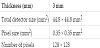
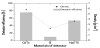
The PID 350CdTe semiconductor detector was designed using GATE in this study. The geometries of the CdTe semiconductor detector are shown in Table 1. The detection efficiency and density of a 3 mm-thick CdTe semiconductor, Si semiconductor andNaI(TI) scintillation detector at gamma ray energy of 140 keV are shown in Figure 1. The 3 mm-thick CdTe semiconductor detector was 76% and 7.6 and 1.4 times higher than that of Si semiconductor and NaI(TI) scintillation detectors, respectively. Accordingly, CdTe semiconductor detector has high efficiency in comparison with other detectors due to high density and high atomic number.
The pinhole collimator is cone-shaped with single aperture. In addition, this collimator is generally consisted of lead or tungsten material due to low cost and appropriate stiffness. Recently, there have been many researches about other collimator materials such as gold and uranium but they have problems with manufacturing and are not commonly available due to relatively high cost [21-22]. Thus, we choose the tungsten material for pinhole collimator in this study. Figure 2 illustrates the schematic diagram of pinhole collimator in simulation. The spatial resolution (Rsystem) and geometric efficiency (g) in the pinhole gamma camera system was calculated by the following formulas [23]:
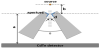
where Rintrinsic is intrinsic resolution of the detector (0.35 mm in this study),Rcollimator is the collimator resolution, a is the distance from the pinhole aperture to the detector surface, b is source-to-aperture distance, deffective is the effective diameter of pinhole, d is the diameter of pinhole, μ is the linear attenuation coefficient of the pinhole collimator material and θ is the acceptance angle of pinhole aperture.
2.2 Evaluation of image performance
To evaluate the image performance with our pinhole systems, the sensitivity and spatial resolution were estimated. A 99mTc point source was measured to evaluate image performance in this study. The activity of 99mTc point source and scan time is 1 MBq and 900 seconds, respectively. Both the sensitivity and spatial resolution were estimated when pinhole diameter was varied from 0.2 to 2 mm by 0.2 mm increment at step for each magnification factor of 2, 3 and 6. Estimated sensitivity was defined as counts per second per kBq (cps/kBq) and spatial resolution was represented in the full width at half maximum (FWHM) of the point spread function. Then, we plotted trade-off curves that express the relationship of sensitivity and spatial resolution to optimize collimator design with respect to the pinhole diameters and magnification factors.
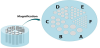
Finally, to confirm overall image performances, we designed a hotrod phantom. The diagram of the phantom is shown Figure 3. The six areas with rods of different diameter were filled with different activity of 99mTc. The phantom was filled with a water solutionof 99mTc.
3. Results and Discussion
The estimated sensitivity and spatial resolution were plotted as a function of the collimator diameters for each magnification factor of 2, 3 and 6 (Figure 4). The results showed that both sensitivity and spatial resolution was improved with increasing magnification factors. The increase of magnification factor means the decrease of source-to-aperture distance with fixed aperture-to-detector distance. It leads to the improvement of geometric efficiency and it makes to the improvement of overall spatial resolution. However, in case of the change of the pinhole diameter, the results of sensitivity and spatial resolution show difference tendency. When increasing pinhole diameter, sensitivity was improved but spatial resolution was aggravated. By using a larger pinhole diameter, we can improve geometric efficiency with larger effective diameter. On the other hand, the larger effective diameter makes to the poorer collimator resolution. Thus, trade-off between sensitivity and spatial resolution is very important for determination of pinhole diameter.
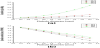
In the most gamma camera system using collimator, it is difficult simultaneously to obtain both high sensitivity and high spatial resolution. For example, in cardiac study in gamma camera system, the acquisition time generally needs to 20 minutes. Due to comparatively long acquisition time, we need to choice high sensitivity collimator. However, when we used high sensitivity collimator, deterioration of spatial resolution must occur. For this reason, we plotted tradeoff curve between sensitivity and spatial resolution to find the best optimal pinhole design. In Figure 5, the trade-off curves of sensitivity and spatial resolution as a function of the pinhole diameter while magnification factor fixed was shown.
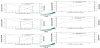
Compared to the each graph in Figure 5, regardless of changes in magnification factor, we found that the optimal pinhole diameter was approximately 1.4mm.
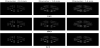
The images of hot-rod phantomto confirm overall image performance were shown in Figure 6. We obtained phantom images with pinhole diameters of 0.4, 1.0 and 1.6 mm and magnification factors of 2, 3 and 6. We found that all rods with diameter of 2.1, 1.8, 1.5, 1.2and 0.85 mm could resolve at all magnification factors and pinhole diameter of 0.4 mm. The 0.5mm rods were certainly resolved when magnification factor and the pinhole diameter were 6 and 0.4, respectively. According to the phantom results, the spatial resolution corresponds to between our proposed system and phantom image.
4. Conclusion
In this study, we designed pinhole gamma camera system with CdTe semiconductor detector using a GATE simulation and evaluated its image performance. The sensitivity and spatial resolution were estimated to evaluate image performance with verifying magnification factors and pinhole diameters. To compromise between sensitivity and spatial resolution, trade-off curve was plotted and we found the optimal pinhole diameter. Finally, we validated overall image performance using a hot-rod phantom. As a result, when pinhole diameter of approximately 1.4 mm is used in all magnification factors, we confirmed that our proposed system acquired the best image performance with reasonable value between the sensitivity and spatial resolution.
Competing Interests
The authors declare that they have no competing interests.
Author Contributions
All the authors substantially contributed to the literature review, drafting the manuscript and approve the final version of the manuscript.
Acknowledgments
This paper was supported by Eulji University in 2016.