


Although the sternotomy incision for cardiac surgery is associated with excellent clinical outcomes sternal instability is reported to occur in a small but significant number of patients (1% to 8%) globally [1- 3]. The literature also suggests that this incidence is a conservative estimate as it only accounts for those patients that are identified in the medical records to have sternal infection or who return to theatre for rewiring [1,4,5]. Sternal instability is described as abnormal motion of the sternum due to bony fracture of the sternum, or disruption of the sternal wires inserted to re-attach the surgically divided sternum [6]. Separation of the sternal halves may be total, involving the entire sternum, or partial, being limited to a portion of the sternum. This result in pain, impaired function and may progress if not diagnosed and managed early to infection and sternal dehiscence [1,2,5,7,8]. Sternal infection, dehiscence and mediastinitis are associated with significant morbidity, mortality and cost of care [1,2,5,7,8]. The risk factors for sternal instability are reported to include pathologic conditions that affect bone healing (diabetes, obesity, COPD), gender (female) or after extensive sternal devascularization following bilateral mammary artery grafting or redo sternotomy [4,9]. It may also develop secondary to prolonged mechanical ventilation (> 24 hours), ace inhibitor medications, or inappropriate forceful unilateral arm activities (falls) [10,11]. This is of relevance in a patient population who are predominantly elderly and increasingly presenting with multiple comorbidities and conditions that compromise bone and wound healing [1,5,12].
Early diagnosis of sternal instability is imperative to arrest the clinical sequelae to sternal infection or mediastinitis [1,5]. Computed Tomography and radiographs of the chest wall has been reported to have low sensitivity and accuracy in the diagnosis of postoperative sternal complications and sternal fractures respectively [3,13]. Real-time ultrasound imaging is a non-invasive, practical and feasible clinical measurement tool [14]. Measures of sternal separation and motion following a sternotomy by Ultrasound have demonstrated validity and excellent test-retest reliability with intra-class correlation coefficients ranging from 0.90 to 0.93 [2,8,14].
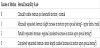
A recent web survey of current practice revealed that 10% of healthcare professionals physically assess the sternum for stability and 20% routinely document subjective patient reporting of symptoms of pain/discomfort instability as part of postoperative evaluation of patients following cardiac surgery [15]. The Sternal Instability Scale (SIS) is a clinical physical assessment tool that aims to assess the stability of the sternum and assign a corresponding grade to the findings of examination (Table 1). It is based on a 4-point scale that is anchored by a grade of 0 that corresponds to a clinically stable sternum with no detectable motion or separation of the sternal edges and a grade 3 that corresponds to a completely separated sternum with marked increased motion or separation of the sternal edges [16]. Previous research has shown that the originally developed 5-point SIS had demonstrated excellent inter and intra-rater reliability, with intraclass correlation coefficients of 0.97 and 0.98 respectively in patients following a median sternotomy with persistent instability [16]. It has demonstrated excellent inter and intra-rater reliability, with intraclass correlation coefficients of 0.97 and 0.98 respectively [16]. The SIS was modified to a 4-point scale to facilitate its compatibility with observations made with real-time ultrasound imaging and the descriptions in the literature that related to regional/partial and complete sternal instability [17]. The validity and reliability of this modified scale had not been investigated in patients following a conventional median sternotomy for cardiac surgery.
The aim of this study was to investigate the validity and reliability of the modified 4 point SIS in patients following cardiac surgery and conventional median sternotomy.
1. Methods
1.1 Study design and Participants
This was a blinded, randomized within-participant experimental study.
1.2 Inclusion criteria
Adult participants undergoing cardiac surgery (Coronary artery revascularization and //or valve surgery via a median sternotomy at a large teaching hospital in Canberra, Australia were invited to participate in this study. Exclusion criteria included patients who were under the age of 18 years; demonstrated impaired cognition or were in a confused state; had received previous chest radiotherapy; and did not comprehend or read the English language. The protocol for this pilot study received institutional ethics approval (Access ID 7736). Written informed consent was obtained from each participant.
All patients received standard post-operative care whilst they were inpatients in hospital and were asked to follow sternal precautions for 4 weeks postoperatively. Sternal precautions included a limitation of 5 kgs when lifting and encouragement to use both arms within the limits of pain and discomfort for safety. Forty participants who had a median stenotomy for cardiac surgery and were a minimum of three weeks post operation that were about to commence cardiac rehabilitation as outpatients were invited to participate in the study. In addition 20 participants who had persistent sternal instability diagnosed by real-time ultrasound imaging and were attending outpatient physiotherapy for conservative management were also included in the cohort of this study. The ultrasound images were captured with a SonoSite M-Turbo device (SonoSite Australasia Pty Ltd, New South Wales, Australia) using a linear array transducer (15- 16 MHz). This transducer has the capacity to scan to a depth of 6 cm below the surface of the skin.
2. Surgical Technique
Each patient underwent sternal closure using cerclage wires that were applied in a figure of 8 or as single wires with 12-gauge stainless steel. Use of bone wax or other hemostatic agents was not documented in all cases.
2.1 Assessors
The assessors included select medical and health professional staff that were involved in the postoperative care of patients following a median sternotomy for cardiac surgery. One cardiologist, one cardiac surgery registrar, one cardiothoracic surgeon, two physiotherapists, two cardiac rehabilitation nurses and one exercise specialist acted as assessors in this study.
2.2 Procedure
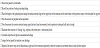
All assessors attended one 30-minute training session that was facilitated by the chief investigator on physical examination and palpation of the sternal separation and excessive movement using the SIS. A set of written guidelines that accompany the SIS was issued to all assessors. This included a checklist to facilitate standardization of assessment (Table 2). Each assessor was blinded from data pertaining to all participants. They were also not known to the participants and had at no time delivered care or treatment to the participants with the exception of medical personnel. Each participant was assigned a number from 1-60 and each assessor received a computer generated random order of numbers to guide the order of assessment. Group assignment was therefore concealed from the assessors. Assessment took place on two occasions that were one week apart with a total of 30 participants being tested by each assessor during one session. Assessors were not provided with any information pertaining to the participant at the time of assessment and were instructed to only assess the sternum manually and record their findings as a single grade on the data sheet provided. A period of ten minutes was allowed for each assessment. Each completed participant data sheet was placed in an envelope and collected by an administrative officer for entry on the summative data sheet at the end of each assessment session. The chief investigator was blinded from the results. The same procedure was followed one week later to obtain the second set of measures. Additionally, Cohen's κ (Fleiss Kappa) was used to determine if there was agreement between the 8 assessors for the SIS scale with the interpretation as follows: values ≤ 0 indicating no agreement; 0.01- 0.20 as none to slight; 0.21-0.40 as fair; 0.41-0.60 as moderate; 0.61- 0.80 as substantial, and 0.81-1.00 as almost perfect agreement [18].
3. Results
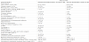
The demographic profile of the two cohorts is presented in Table 3.
4. Validity
4.1 Criterion validity
All patients were assessed by real-time Ultrasound to screen for sternal instability prior to physical examination of the sternum and application of the SIS. There was significant correlation of US measures with manual assessment with 95% exact agreement on the SIS scale.

4.2 Discriminative validity
Patients diagnosed with sternal instability were rated higher on the SIS compared to those who did not have an unstable sternum on the SIS. The mean difference on the SIS between those individuals with sternal instability and those who had a stable sternum was 2.35 SIS grade points on the SIS.

5. Reliability
5.1 Intra-rater reliability
The intra-rater reliability of the mean SIS scores for all 8 assessors ranged from ICC (3,1) 0.92 to 0.99.
5.2 Inter-observer reliability
The inter-observer reliability of the SIS scores for all 8 assessors over the two occasions of testing yielded an ICC (2,1) mean value of 0.88. There was fair to moderate agreement between the 8 assessors with Cohen κ = 0.36 (95% CI 0.31, 0.41), p< .0005.
5.3 Incidental observations
Twenty participants with persistent sternal instability 6 weeks post operation to ten years since onset participated in this study. Real-time Ultrasound assessment of the sternum revealed a range of 6mm to 45mm separation of the sternal edges. All participants with sternal instability reported symptoms of pain or discomfort that impacted on activities of daily living in particular tasks that involved upper limb movements such as driving, transfers from bed to sitting and sleeping. Two participants with prolonged sternal instability and nonunion (male: 5 years postoperative; female: 10 years postoperative) demonstrated a connective tissue/scar bridging the sternal halves on Ultrasound.
6. Discussion
Sternal instability accompanied by wound infection or alone is a postoperative complication that exists in a small but significant number of patients following cardiac surgery via a median sternotomy [1,3,7]. The early recognition and diagnosis of sternal complications is imperative to ensure timely management that arrests the clinical sequelae to deep infection and mediastinitis. Real-time ultrasound is a valid and reliable imaging tool for the diagnosis and quantification of sternal instability and separation respectively. However, in the absence of Ultrasound physical assessment of the sternal edges for separation and excessive motion accompanied and reference to the SIS yields valid results and reporting of sternal instability. More specifically the SIS has excellent criterion validity as there was a 95% exact agreement between a diagnosis of sternal instability made by real-time Ultrasound and that made by physical examination of the sternum using the SIS (grade ≥ 2). With respect to discriminative validity and the feasibility of the SIS to predict differences between patients diagnosed with (grade ≥ 2) and without sternal instability (Grade 0) there was a significant difference between group means that equated to 2.35 SIS grade points higher on the SIS respectively.
In addition the physical examination of the sternum and screening of sternal instability using the SIS yielded excellent Intra-Rater reliability with estimates ranging from 0.92 to 0.99 for the 8 raters who were representative of medical and health professional personnel that are involved in the postoperative care of cardiac surgery patients selected for this this study. The Inter-rater reliability for all 8 raters over the two occasions of testing yielded an ICC (2,1) mean value of 0.88 which equates to excellent reliability [17].
The SIS provides a consistent and standardised method of physical assessment to screen and monitor sternal stability. It is a non-invasive, practical manual clinical assessment tool that requires minimal training and can be administered by health professionals in the acute and community settings for the purposes of screening and diagnosis; monitoring progress following conservative or surgical management. More importantly it facilitates standardised and consistent reporting amongst health professionals and prompts early referral to medical and surgical practitioners for review and timely intervention.
Future research should target development of normative SIS grades following conventional sternotomy in patients at high and low risk of sternal complications; and evaluation of reliability and validity of this scale in varying methods of sternal closure.
7. Conclusion
The SIS is a valid and reliable clinical scale that facilitates the assessment, screening and diagnosis of sternal instability following cardiac surgery via a median sternotomy.
Competing Interests
The authors declare that they have no competing interests.
Acknowledgments
The authors would like to thank the respective staff in the Cardiac Surgery, Cardiac Rehabilitation, Cardiology and Physiotherapy departments at Canberra Hospital (ACT, Australia) and the participants who took part in this study.