


1. Introduction
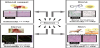
Because of their structural rigidity and compositional resemblance, ceramic biomaterials or bioceramics have been studied and developed for replacement and reconstruction of hard tissues of bones and teeth. Additionally, considering their potentiality of excellent compatibility to skins and blood vessels, bioceramics have currently been also expected to extend to repair of soft tissues. However, as-prepared surface properties of conventional bioceramic products are actually insufficient for fulfilling such biomedical multi-purposes. As a result, we are keenly required to develop novel bio-interface engineering, with which surrounding media such as organic and inorganic constituents, and living tissues and cells in vivo could be controlled, in order to realize such extensive goals of clinical applications. For this purpose, we propose the development of vector materials [1], which can influence a force on and manipulate a desired spot of wounded tissues under a certain circumstance of a quasi-closed system such as human body (Figure 1). It is, however, needless to say that not only improvement of materials processing and surface coating technology, but also more fundamental issues such as control of surface free energy chemical stability should be included.
2. Vector Biomaterials
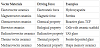
Magnets are therefore typical vector materials after the definition, because magnets irradiate magnetic field from the north to the south poles and affect a force on a substance placed between the two poles. A radioactive material for cancer therapy is another candidate of vector material because they can individually irradiate a force to its surrounding. Both bioglass and β-tricalcium phosphate (TCP), which gradually dissolve into a body and release chemical constituents or artificial chemicals, resulting in good bone conductivity. These materials fall to chemicovector materials. Virus-vector can be said as one of vector materials, and DNA- and drug-carrying proteins also fall into this category. We summarize these materials and effects as Table 1, giving the classification of the family of vectors ceramics on the basis of the mechanisms to drive the effects. Some functional materials are under development for vector materials.
We have been developing the surface modification for a vector material by means of electrical polarization of bioceramics. Based on the finding of remarkable effects of polarized electrets, electrovector effects [1] are employed as a concept exceeding bioactivity. Conventionally polarization has been applied mainly to ferroelectric or piezoelectric ceramics, in which spontaneous dipoles are predominantly arranged in a dc field of polarization. In our study of bioceramics and bioglasses, polarization has been proved to take place due to ionic displacement [2] to a rather long range. Electrically polarized materials are classified as electrets, named after magnets, in physics, and bioceramic electrets are called vector materials. Electrically polarized ceramic hydroxyapatite was, above all, experimentally proved an excellent vector material, which was effective for manipulation of the surrounding constituents including cells and tissues in vivo. Other members of calcium phosphates family show also outstanding performance as polarized materials. This report briefly presents the effects of hydroxyapatite electrets on the basis of the current results. Based on the results on hydroxyapatite, the novel bio-interface engineering is proposed for development of biomaterials.
3. Fundamentals of Hydroxyapatite Electrets
Hydroxyapatite exhibits important and interesting electrical properties. The electrical properties of hydroxyapatite arise from its stable lattice hydroxide ions surrounded with calcium ions in the crystal structure. At higher temperatures, hydroxyapatite ceramics show a rather higher conductivity of 10-7 down to 10-4 Scm-1 [3]. Besides, it was demonstrated through a concentration cell measurement of the electromotive force generated using a hydrogen concentration cell of hydroxyapatite ceramic that the mass transport takes place due to the migration of protons [4]. The recent studies by the thermally stimulated current method showed that hydroxyapatite ceramics were polarizable at a temperature higher than the monoclinic-to-hexagonal phase transition temperature (500 K). The dielectric measurements also exhibit dielectric characteristics around polarization temperature of 300 - 500 K [5]. Polarization takes place due to the reorientation of the lattice OH- ions along the c-axis. Relaxation or depolarization occurs only when the strain induced by electric stress is thermally released. Using the measured current-temperature curve, we can evaluate the stored energy in hydroxyapatite giving a few tens of μCcm-2.
The first vector effect was observed on the crystal growth on hydroxyapatite ceramic electrets: Although slow crystallization took place dispersedly on non-polarized hydroxyapatite, large crystals of 1-4 μm in diameter covered the negatively-charged surface of the electret after an immersion in 1.5SBF for only 12 h. Higher field strength gave rise to faster crystal growth; sizable agglomerated crystals of 10 μm in diameter were observed in some spots on polarized hydroxyapatite under 1 kVcm-1. The accelerated crystal growth mentioned above was observed on the negatively-charged surface, whereas no crystal growth ever took place on the positively-charged surface even after 3-day immersion in 1.5SBF. The crystal growth was dependent upon the dc field strength, temperature and time for polarization.
Recent development for biomedical applications is summarized in Figure 2, which shows that bioceramic electrets have excellent reconstructive effects not only on bones and teeth [6], but on soft tissues such as wound skins, blood vessels, and nerve [7-9]. The ongoing biological, chemical, and physical analyses will contribute to novel bio-interface engineering; advancements are very recently reported on new calcium phosphate electret [10] and bio-interfacial analysis [11].

4. Summary
Because of the growing number of aged people, the need of multifunctional biomaterials will be more growing in the near future. Under such circumstances, it is reasonably expected that some of potential vector ceramics will play an important center role in clinical treatment.
Competing Interests
The authors declare that they have no competing interests.