


Data emerging from the literature show how diverticulitis may turn out to become complicated in about 25% of cases (abscess, fistulization, bleeding, obstruction with or without stricture, peritonitis up to sepsis), of which approximately 15% need surgery [1]. Generally, no previous symptoms are related to the underlying diverticular disease [1].
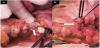
A 57-year-old woman was admitted to our Unit for pneumaturia and fecaluriaassociated with fever (38-39°C), appeared in the three days prior to the visit. An anamnestic history did not reveal pastdiagnosis of diverticular disease. Furthermore, there was no personal or family history of inflammatory bowel disease, no prior findings of colon cancer,except fora previoushysterectomyperformed for a benign disease. On admission,increased white blood cell count (17,23/mm3; neutrophils 87,8%) and index of inflammation (CRP 5.3 mg/dL) were present. On physical examination, the abdomen was poorly treatable with signs of mild peritoneal reaction. The patient underwent an abdominal computed tomography with contrast agent showing signs of sigmoid diverticulitis and gas in the bladder. A fistulography revealed a colovescical fistula. A one-step laparotomywas led in order to achieve a better visualization of the surgical field. At the beginning of the procedure, the sigmoid colon appeared strongly adherent to the bladder and the location of the fistula was observed (Figure 1). After adhesiolysis, a left hemicolectomy associated with repair of the bladder’s wall defect were performed. Bowel rest, parenteral nutrition and intravenous broad-spectrum antibiotic therapy were started. The patient recovered well and was discharged 10 days after in good condition.
In case of diverticulitis, the incidence of fistulas isabout 4-20% with a prevalence of thecolovesical ones (CVF) [2], more frequently occurring in males, as in females the uterus is located between thecolon and bladder.This finding is supported by the observationthat the majority of women with fistulas (colovesical or colovaginal) underwent a prior hysterectomy [3]. Recently, Miyaso et al. reported10 cases of CVFs, caused by sigmoid diverticulitis, showing how fecaluria and pneumaturia were present in 40% and 30%, respectively, as clinical presentation [4]. In conclusion, CVF due to sigmoid diverticulitis is a relatively rare disease and early surgical treatment is the best option.
Competing Interests
The authors declare that they have no competing interests.