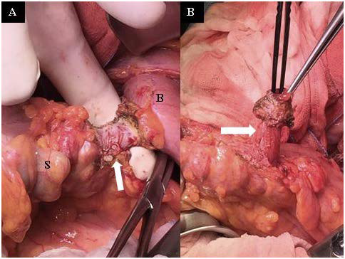
Figure 1: Surgical view:A) the sigmoid colon (S) and the bladder (B) were adhered to each other, and it was possible to identify the penetrating sigmoid diverticula into the bladder (white arrow); B) forceps showed the fistulazing diverticula opened in the bladder (white arrow).