Profile
International Journal of Gastroenterology Disorders & Therapy Volume 4 (2017), Article ID 4:IJGDT-125, 4 pages
https://doi.org/10.15344/2393-8498/2017/125
Case Report
Metastasis of the Proximal Jejunum from Cervical Carcinoma: A Case Report and Literature Review

Lin Li1, Guibin Yang1, Jianwei Zheng2, Chunbo Kang2, Jie Wang1, Yuanmin Zhu1* and Jun Qu2*

1Department of Gastroenterology, Peking University Aerospace Center Hospital, Yu-Quan Road, Beijing, China
2Department of General Surgery, Peking University Aerospace Center Hospital, Yu-Quan Road, Beijing, China
Dr. Yuanmin Zhu, Department of Gastroenterology, Peking University Aerospace Center Hospital, #15 Yu-Quan Road, Hai-Dian District, Beijing 100049, China; E-mail: zhuyuanmin@sina.com
Dr. Jun Qu, Department of General Surgery, Peking University Aerospace Center Hospital, #15 Yu-Quan Road, Hai-Dian District, Beijing 100049, China; E-mail: qujun@asch.net.cn
15 December 2016; 07 February 2017; 09 February 2017
Li L, Yang G, Zheng J, Kang C, Wang J, et al. (2017) Metastasis of the Proximal Jejunum from Cervical Carcinoma: A Case Report and Literature Review. Int J Gastroenterol Disord Ther 4: 125. doi: http://dx.doi.org/10.15344/2393-8498/2017/125

Abstract

Metastatic neoplasm of the small intestine is uncommonly encountered. We herein reported a case of 66-year-old female patient with a history of cervical carcinoma presented as a spell of digestive symptoms. Imaging examination revealed disseminated disease involving her proximal jejunum. Endoscopic biopsy was performed showing metastatic squamous cell carcinoma with the same characteristics of cervical lesion. Although the clinical presentation of enteral metastases is extremely rare, awareness of such metastatic pattern of cervical cancer is necessary for diagnostic practices and therapeutic decisions.


1. Introduction

Malignant tumors of the small bowel are uncommon tumor accounting for only 1-5% of all gastrointestinal tract malignancies [1]. Histologically, adenocarcinoma is most frequently involved, followed by mesenchymoma, lymphoma, leiomyosarcoma, and metastatic carcinoma constituting the minority. Generally, gastrointestinal metastases are rare and asymptomatic, occurring in advanced tumor stages, and usually accompanied by poor prognosis. Most of the metastatic intestinal tumor originates from lung, melanoma, choriocarcinoma and breast, but with very few cases of cervical carcinoma involvement.

Herein, we share a rare case of metastasis of the proximal jejunum from cervical carcinoma presenting as postprandial nausea and vomiting. The diagnostic procession is depicted and a brief review of such metastatic diseases is provided.

2. Case Presentation

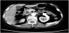
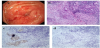
A 66-year-old female were hospitalized with severe gastrointestinal symptoms. Patient had complaints of appetite loss, postprandial nausea, intermittent vomiting and abdominal bloating. She was diagnosed with stage IIb squamous cell carcinoma of cervix three years ago and received concurrent chemotherapy and radiation treatment. She had undergone local radiotherapy 1 month previously for pelvic lymph node metastasis. An abdominal CT revealed the presence of prominent mural thickening with proximally luminal dilatation involving duodenojejunal junction (Figure 1). The subsequent upper endoscopy identified a protuberant lesion with luminal narrowing located at proximal jejunum about 5 cm distal to the Treitz ligament. The mucous membranes appeared to uneven, redness and erosion (Figure 2a). Biopsies were performed and pathological examination showed solid nests of poorly-differentiated cancer cell in mucosa and submucosa emerging from chronically inflamed intestinal mucosa (Figure 2b). Additional immunohistochemistry supported the diagnosis of squamous cell carcinoma based on the results of histochemical staining: P63 (+), CK5/6 (+), P40 (+), CK7 (-), and CK20 (-) (Figure 2c, d). In view of these data, the lesion was considered to be cervical carcinoma and underwent self-expanding metal stent insertion via endoscopy. On postoperative examination, intestinal obstruction was relieved (Figure 3). 5 days after decompression and supportive management, the patient was asymptomatic for the metastasis and discharged. At present, she is on a regular follow-up.

figure 1
Figure 1: CT scan showed the presence of mural thickening and luminal dilatation involving the jejunum.
figure 2
Figure 2: Endoscopic and histopathological images of the enteral metastasis. (a) Endoscopic view of the jejunal tumor, (b) HE staining showed inflamed mucosa with squamous islands (100×), (c, d) Immunohistochemical photomicrographs showed the tumor cells were positive for P63 (c, 100×) and negative for CK7 (d, 100×).
figure 3
Figure 3: Re-examination of abdomen x-ray after self-expanding metal stent placement.

3. Discussion

Cervical neoplasia is one of the commonest gynecologic malignancy seen in China accounting for approximately 29000 deaths per year [2]. About 60-80% of cervical neoplasia is squamous cell carcinoma, 5-15% is adenocarcinoma and less 10% consists of adenosquamous carcinoma [3]. In essence, squamous cell carcinoma of cervix tends to spread locally, commonly restricted to pelvic structure. Lymphatic spread principally accounts for the metastatic lesions beyond the pelvis and has the capacity to skip to distant nodes generating isolated lesions anywhere in the body. The most frequent sites of metastatic lesions include lung (21%), bone (16%) liver (4%) and gastrointestinal tract (4%) [4,5]. Intestinal metastases from cervical tumor are mostly at rectosigmoid portion because of local extension, while upper intestinal metastasis is exceedingly uncommon. It remains puzzling why small bowel metastases are such rare events. The potential antitumor mechanism involves abundant mucosal immune protection, and strong self-renewal capability [6,7].

Our search of the literature found 12 articles reporting on cervical carcinoma patient with biopsy-proven metastasis in small intestine so far. There are 6 cases of duodenal metastases [8-13], 2 cases of jejunal metastases [14,15], and 4 cases of ileum involvement [16-19]. A comprehensive summary of such cases is provided in Table 1. The majority of these patients are found to have intestinal metastasis within the first 2 years after diagnosis of the primary cervix tumor. The metastases can cause bowel obstruction, bleeding and epigastric pain. On endoscopy, tumor lesions are presented as polypoid mass, nodules or submucosal mass with ulceration. Cancer patients with small bowel involvement usually have a poor prognosis due to delayed diagnosis and high malignant degree. Given the rarity of the condition, there remains therapeutic uncertainty and limited data to guide treatment decisions. In this context, treatments could be chosen based on performance status of the patients. As in our case, palliative self-expanding metal stent implantation is the best way to relieve symptoms and improve survival quality. Metal stent insertion has been reported as a promising palliative treatment for patients with advanced malignancies causing gastrointestinal obstruction. The feasibility, efficacy and safety of this technique have been evaluated in few studies [20,21]. Bessoud et al. reported on 72 patients with malignant small bowel obstructions who underwent metal stent insertion. The technical success rate was 97% while the complication rate was 17%. The most common complications include stent migration, stent fracture, and intestinal perforation [22]. Similarly, Kim et al evaluated the complications of metal stent insertion in 213 patients with malignant gastroduodenal obstruction. A total of 45 (21%, 45/213) complications occurred in the first year after stent placement. Tumor overgrowth, stent collapse, and stent migration were the main complications affected the prognosis [23]. In our case, there is no short-term complication and the patient will be followed up.
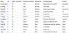
table 1
Table 1: The intestinal metastases from carcinoma of cervix reported in the literature.
NR-not reported.

4. Conclusion

In conclusion, despite its rarity, the possibility of metastasis to jejunum or other gastrointestinal tract involvement should be kept in mind. Besides, nausea, vomiting and appetite loss are common symptoms of cancer patients in advanced stage especially during the course of chemo radiotherapy. Failure to consider intestinal metastasis may result in a delay in diagnosis. Physicians should be aware of the varied metastatic lesions and perform endoscopic examination of necessary.

Competing Interests

The authors declare that they have no competing interests.


References

  1. Torres M, Matta E, Chinea B, Dueno MI, Martinez-Souss J, et al. (2003) Malignant tumors of the small intestine. J Clinical Gastroenterol 37: 372- 380. View
  2. Di J, Rutherford S, Chu C (2015) Review of the Cervical Cancer Burden and Population-Based Cervical Cancer Screening in China. Asian Pac J Cancer Prev 16: 7401-7407. View
  3. Cheah PL, Looi LM (1999) Carcinoma of the uterine cervix: a review of its pathology and commentary on the problem in Malaysians. Malays J Pathol 21: 1-15. View
  4. Fagundes H, Perez CA, Grigsby PW, Lockett MA (1992) Distant metastases after irradiation alone in carcinoma of the uterine cervix. Int J Radiat Oncol Biol Phys 24: 197-204. View
  5. Bhandari V, Kausar M, Naik A, Batra M (2016) Unusual Metastasis from Carcinoma Cervix. J Obstet Gynaecol India 66: 358-362. View
  6. Loualidi A, Spooren PF, Grubben MJ, Blomjous CE, Goey SH (2004) Duodenal metastasis: an uncommon cause of occult small intestinal bleeding. Neth J Med 62: 201-205. View
  7. Schottenfeld D, Beebe-Dimmer JL, Vigneau FD (2009) The epidemiology and pathogenesis of neoplasia in the small intestine. Ann Epidemiol 19: 58-69. View
  8. Subramanian K, Aroul T, Kuppusamy S, Gunashekaran V (2016) Intra Luminal Metastasis to Duodenum: A Histological Surprise. J Clinical Diagn Res 10: 8-10. View
  9. Chawhan SM, Dani AA, Meshram SA, Randale AA, Kumbhalkar DT (2015) Metastases of squamous cell carcinoma of uterine cervix to duodenum: a case report. J Clinical Diagn Res 9: ED03–ED04. View
  10. Raphael JC, Ram TS, Pavamani S, Choudharie L, Viswanathan PN (2011) Squamous cell carcinoma cervix with metastasis to pyloroduodenal region. J Cancer Res Ther 7: 183-184. View
  11. Kanthan R, Senger JL, Diudea D, Kanthan S (2011) A review of duodenal metastases from squamous cell carcinoma of the cervix presenting as an upper gastrointestinal bleed. World J Surgical Oncology 9: 113. View
  12. Lee TH, Park SH, Lee CK, Lee SH, Chung IK et al. (2011) Ampulla of Vater metastasis from recurrent uterine cervix carcinoma presenting as groove pancreatitis. Gastrointest Endosc 73: 362-363. View
  13. Gurian L, Ireland K, Petty W, Katon R (1981) Carcinoma of the cervix involving the duodenum: case report and review of the literature. J Clin Gastroenterol 3: 291-294. View
  14. Misonou J, Natori T, Aizawa M, Jou B, Tamaki A, et al. (1988) Stage (Ia) cervical cancer recurring 13 years after hysterectomy and causing small intestinal perforation. A case report with a review of the literature. Acta pathologica japonica 38: 225-234. View
  15. Onal C, Nursal GN, Torer N, Kayaselcuk F (2015) Isolated jejunal metastasis in a patient with cervical cancer: A case report. Reports of practical oncology and radiotherapy. Rep Pract Oncol Radiother. 20: 239- 242. View
  16. Mathur SK, Pandya GP. Solitary metastatic malignant stricture of the ileum: a rare cause of small bowel obstruction (a case report). J Postgrad Med 30: 186-188. View
  17. Qiu H, Yuan L, Ou Y, Zhu Y, Xie C, et al. (2015) Small intestine metastasis from cervical cancer with acute abdomen: A case report. Oncol Lett 9:187- 190. View
  18. Sugimoto T, Mike M, Abe M, Kano N (2013) Small bowel metastasis of uterine cervical adenocarcinoma. BMJ Case Rep 2013: 007896. View
  19. Yu X, Wang Z, Zhang Z, Liu Y, Huang J (2016) Postoperation of cervical cancer with intestine metastasis: a case report and literature review. World J Surg Oncol 14. View
  20. Yim HB, Jacobson BC, Saltzman JR, Johannes RS, Bounds BC, et al. (2001) Clinical outcome of the use of enteral stents for palliation of patients with malignant upper GI obstruction. Gastrointest Endosc 53: 329-332. View
  21. Nakahara K, Okuse C, Matsumoto N, Suetani K, Morita R, et al. (2015) Enteral metallic stenting by balloon enteroscopy for obstruction of surgically reconstructed intestine. World J Gastroenterol 21: 7589-7593. View
  22. Bessoud B, de Baere T, Denys A, Kuoch V, Ducreux M, et al. (2005) Malignant gastroduodenal obstruction: palliation with self-expanding metallic stents. J Vasc Interv Radiol 16: 247-253. View
  23. Kim JH, Song HY, Shin JH, Choi E, Kim TW, et al. (2007) Metallic stent placement in the palliative treatment of malignant gastroduodenal obstructions: prospective evaluation of results and factors influencing outcome in 213 patients. Gastrointest Endosc 66: 256-264. View