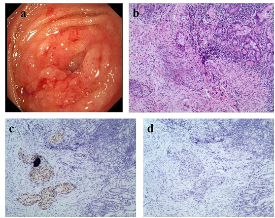
Figure 2: Endoscopic and histopathological images of the enteral metastasis. (a) Endoscopic view of the jejunal tumor, (b) HE staining showed inflamed mucosa with squamous islands (100×), (c, d) Immunohistochemical photomicrographs showed the tumor cells were positive for P63 (c, 100×) and negative for CK7 (d, 100×).