


1. Introduction
Inflammatory pseudotumor (IPT) also known as inflammatory myofibroblastic tumor is a rare tumor-like lesion of unknown cause. It may arise in nearly every site of the body (e.g. breast, kidney, thyroid, salivary glands, larynx, spleen, stomach, brain, omentum, mesentery, orbit), but most commonly develops in the lung and the liver [1-8]. The first description of hepatic localization appeared in 1953 [9]. In most cases it affects adults, but in Asia, the hepatic IPT occurs often in children and young adult persons [2,6]. IPT usually presents as an asymptomatic [5], slowly growing, unilateral mass accompanied by unspecific symptoms like fever, malaise, abdominal pain and rarely vomiting, jaundice and weight loss [3,4,6,7,10]. It typically forms the solitary large (maximum dimension range from 2 to 22 cm) mass and accounts for 0.4% of all focal liver lesions [2,7]. Less often multifocal lesions have been described [2,7,11-13]. Majority cases are located in the right lobe. Radiological findings of the IPT are nonspecific but benign histopathological features constitute the hallmark of this lesion [12-14,16,17].
The aim of the description was to present a new case of hepatic inflammatory pseudotumor in patients with type 2 diabetes.
2. Case Report
A 65-year-old woman was admitted to the 1st Department of General Surgery of Medical University of Lublin (Lublin, Poland) with weakness, increased temperature and a liver mass discovered incidentally on the ultrasound scan. The right upper abdominal quadrant was tender at palpation. There were no signs of jaundice. Her previous medical history revealed classical cholecystectomy performed due to cholelithiasis about 6 years ago. Abdominal ultrasonography (USG) showed a 50x42x39 mm hypoechogenic, slightly heterogenous lesion located in the right lobe of the liver (segment 4/5). Computer tomography (CT) confirmed presence of irregular, hypoattenuating pathological mass with an irregular contrast enhancement in portal and venous phase in segments 4/5. Higher enhancement was revealed only in arterial phase. Similar subcapsular lesion with poorly visible margins (57x15 mm) was found in segment 7. Mild enlargement of mesenteric and hepatic hilar lymph nodes was observed. The primary radiological diagnosis was hepatic tumour with non-excluding malignancy or abscess but the second lesion was regarded as posttraumatic damage.
The results of laboratory tests were as follows: white blood cell count – 12.360/μL (normal range 4.000-10.500/μL), neutrophils – 76.2% (normal range 45-70%), lymphocytes – 15.7% (normal range 20-45%), monocytes – 5.3% (normal range 3.0-8.0%), red blood cell count – 3.53x10⁶/μL (normal range 3.8-5.2x x106/μL), serum hemoglobin concentration – 10.4 g/dL (normal range 12.0-16.0 g/dL); haematocrit – 31.2 % (normal range 37.0-47.0), and serum C-reactive protein – 230.18 mg/l (normal range <5.000 mg/l). The laboratory data showed moderately elevated liver enzyme: aspartate aminotransferase (AST) – 42 U/L (normal<34 U/L) and alanine aminotransferase (ALT) – 47 U/L (normal <31 U/L).
Other results of biochemical analysis of the blood including serum platelet count and levels of serum total bilirubin, alkaline phosphatase (ALP), amylase and lipase were within normal limits. Laboratory parameters of renal function and serum level of tumor markers (α-fetoprotein) – remained within normal range. Hepatitis B virus antigen and hepatitis C virus antibody were negative. Furthermore, the patient suffered from diabetes type-2 which was controlled by diet and Metformin administration. Her fasting plasma glucose level was 99mg/dL.
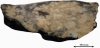
An initial clinical diagnosis of hepatic tumor was made. The patient was qualified for urgent laparotomy with frozen section examination. Samples taken during surgical operation revealed prominent inflammatory process with numerous plasma cells, lymphocytes and foam cells with accompanied fibrosis. In the subsequent step, the patient underwent resection of the whole hepatic lesion. The ultimate surgical specimen sized a 6.5x5.5x3,0 cm contained an ill-defined, solid, firm, light-brown lesion with white-gray areas a 5x2,5x2 cm in dimentions (Figure 1). After fixation in 10% buffered formalin, many samples were taken for histopathological examination and then routinely processed into paraffin blocks. The slides were stained with hematoxylin and eosin (HE), mucycarmine, periodic acid Shiff (PAS), van Gieson, Prussian blue and Shikata’s orcein method for hepatitis B surface antigen (HBsAg). Furthermore, immunohistochmical reactions with smooth muscle actin (SMA), vimentin, CD34, CD117, ALK1, CD68, CD138, CD45 and Ki67 were also performed using reagents from Dako (Denmark) according to manufacturer directions.
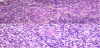
Microscopically, the lesion was composed of the anastomosing fascicles of spindle cells intermingled with a variety of inflammatory cells including lymphocytes (CD45+), plasma cells (CD138+), different types of macrophages i.e. foamy histiocytes and multinucleated giant (CD68+) and few neutrophils, occasionally surrounded by collagen bundles (Figure 2A and Figure 2B). An immunohistochemical analysis revealed some SMA- (Figure 3) and vimentin-positive spindle cells, but they were negative for ALK1, CD34 and CD117. Proliferative index (Ki67) was low (5-7%). Staining for HBsAg was negative. Resection margin was free of the lesion but some scattered hepatocytes with macrovacuolar steatosis were present. There was no evidence of malignancy. A final histopathological diagnosis of IPT was established.

Follow-up CT performed two weeks after the surgery demonstrated a small fluid-filled cystic space in the postoperative area. But apart from that the postoperative period was uneventful. The patient becomes asymptomatic one year after the surgery.
3. Discussion
IPT is an uncommon hepatic lesion considered as a benign condition usually with good prognosis, however, aggressive behavior has been occasionally reported [3,18]. They may sporadically recur usually as a consequence of incomplete resection [14,18]. Both, clinical and histopathological features of IPT did not predict the aggressive behavior [18].
The etiology and pathogenesis of this entity remain unclear. Many mechanisms and hypothesized etiologic factors like infections, trauma, autoimmunity reactions, radiotherapy, and local reaction to changes in the walls of bile ducts are suggested [1-7,10,13,18-26]. Some microorganisms that access the portal vein including Escherichia coli, Staphylococcus aureus, Klebsiella pneumoniae, Gram-positive cocci and Actinomyces were regarded as causative agents. Viral infections (Epstein-Barr virus [20], hepatitis B and C virus) [10,27] and parasitic infestations (Schistosoma mansoni) may play role in the pathogenesis [18]. Some authors suggested that disturbance of biliary drainage, e.g., due to biliary stones [1,7], recurrent pyogenic cholangitis [4,26] or sclerosing cholangitis is also important [25]. The tumor was also noted in HIV infected persons [24]. IPT is occasionally associated with systemic diseases, mostly of autoimmune background like Crohn disease [19,21], autoimmune pancreatitis [23], as well as diabetes mellitus [7,28], gout, acute myelomonocytic leukemia [29] and rectal gastrointestinal stromal tumor [30]. More recently some genetic rearrangements referring anaplastic lymphoma kinase 1 (ALK1) expression has been reported in IPT [3,7,31-33]. Chromosomal translocation involving chromosome 2p23 upon which the ALK gene is located has been identified in some cases [31-33]. Moreover a fusion of ALK gene with a partner genes such as TPM3 and TPM4 has been demonstrated [33]. Although ALK-positive and ALK-negative tumors are histologically identical, variable immunophenotype support the heterogeneity of IPTs and may indicate a different pathogenetic processes in hepatic IPT [18,31-33]. In presented case immunohistochemical reaction with ALK1 was negative.
The histological appearance of IPT can vary and thus multiple terms were given for this condition, e.g. plasma cell granuloma, inflammatory myofibroblastic tumor, histiocytoma, fibroxanthoma, xanthogranuloma or plasma cell histiocytoma [1-5,10,17,18,31,33]. According to the current WHO classification of the digestive system tumor [2], it is defined as a mesenchymal, benign, non-neoplastic and non-metastasizing mass composed of fibrous tissue and proliferated myofibroblasts with a marked inflammatory infiltration, predominantly of plasma cells. Marked cellular atypia and frequent mitoses are not found in IPTs [2,4,5,7,8]. IPT always consists of a mixture of inflammatory cells and fibrotic tissue but the relative proportion of these components can be variable. Due to microscopic heterogeneity, they were previously classified into three histologic subtypes, e.g. hyalinised sclerosing, xanthogranuloma and plasma cell granuloma [6]. However, recently only two types are distinguished: fibrohistiocytic and lymphoplasmacytic from IgG4-related disease [7,8,11]. Reported case fulfils histological criteria of fibrohistiocytic type, which is more frequent. Zen et al. [8] suggested that IPT may represent the end stage of heterogeneous inflammatory processes in the liver, including cholangitis. Recently Yang et al. [7] performed a retrospective analysis of 114 patients with hepatic IPT and proposed a clinical diagnosis guideline for establishing a correct diagnosis. It's interesting that in their study 30 patients had a history of biliary tract inflammatory disease in the past and 18 suffered from diabetes mellitus. They suggested that both above mentioned conditions are associated with development of IPT in the liver. In the present case two potentially predisposing factors were noted, i.e. cholelithiasis accompanied by chronic cholecystitis in the past medical history which could be regarded as an induction factor and successfully treated type 2 diabetes.
Regardless location IPT may be confused with true neoplasms. Precise diagnosis of lesions is difficult because they are rare conditions and clinical symptoms and laboratory findings are nonspecific. Furthermore, both clinical presentation and radiological images may be suggestive for malignancy [4,11-13,15,16,22,27,30,31]. In the liver IPT has to be distinguished from hepatocellular carcinoma [15,16,27], intrahepatic cholangiocarcinoma [11,13], metastases [12,30] and inflammatory conditions mostly abscesses [17,22]. For this reason IPT is sometimes called ‘the great mimicker” [22]. To avoid inappropriate diagnosis of neoplasm a detailed microscopic examination of the whole tumor with many samples proceeded by biopsy or frozen-section examination is necessary. Although radiographic appearance of the described tumor has not ruled out malignancy, the microscopic features were characteristic. Exclusion of neoplastic process is the primary concern, since IPT can be resolved by conservative therapy within 1-2 months (antibiotics, steroids or nonsteroidal anti-inflammatory drugs) or by surgery with excellent prognosis [11,12,34,35]. Spontaneous regression of hepatic IPT has been reported as well [36].
In conclusion, diagnosis of inflammatory pseudotumor is mostly based on microscopic examination, since its clinical and radiological presentation is frequently not characteristic. Correct diagnosis is essential for proper management.
Competing Interests
The author(s) declare that they have no competing interests.