


1. Introduction
The status of the groin lymph nodes is the most important prognostic factor for patients with vulvar cancer. Selected early vulvar cancers may be amenable to sentinel node biopsy, but many patients will require an inguino-femoral lymphadenectomyin order to adequately treat the groin nodes.
The use of a separate incision approach significantly improved wound healing and decreased post-operative hospital stay, but the long-term problem of lower limb lymphedema remained. Several attempts have been made to try to reduce the risk of lymphedema, including elimination of groin dissection in patients with ‘microinvasive’ vulvar cancer [1], the performance of a superficial inguinal lymphadenectomy [2] and the use of primary groin irradiation. These approaches were shown to increase the incidence of groin recurrence [3-5].
The purpose of this study was to determine the incidence of short and long-term postoperative morbidity of groin node dissection in a large cohort of patients, to investigate causal factors, and to postulate possible strategies to further reduce this morbidity.
2. Materials and Methods
2.1 Study design
A retrospective observational single institutional study.
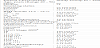
Following ethics approval obtained from the South Eastern Sydney Local Health District Human Research Ethics Committee (Reference Number 15/151), the medical records of 429 consecutive patients treated for primary invasive vulvar cancer at the Royal Hospital for Women in Sydney, between February 1987 and June 2016 were reviewed. Ninety-six patients were excluded as their groins were not surgically treated. The remaining 333 patients underwent either unilateral or bilateral inguino-femoral lymphadenectomy, groin node debulking, or a sentinel node procedure and were included in the analysis. Data retrieved from the medical records included age at diagnosis, body mass index (BMI), smoking status, co-morbidities, disease stage, tumour diameter, histologic type, histologic grade, primary treatment, adjuvant treatment, type of lymph node dissection, number of lymph nodes removed, intra-operative insertion of a groin drain, duration of drain use, post-operative groin wound infection, groin wound dehiscence/breakdown, lymphocyst formation, length of stay and hospital readmission. Follow up data on lymphedema and patient disease status was retrieved from the outpatient clinical files. All patients were staged according to the 2009 International Federation of Gynecology and Obstetrics (FIGO) staging system [6].
Lymphocyst formation was recorded if confirmed by an ultrasonic scan, or if fluid was drained from the groin. Groin wound breakdown was defined as opening of the wound requiring either wound packing, or a negative pressure dressing. Groin wound infection was defined as erythema or a purulent exudate necessitating the use of antibiotics. Chronic lower limb lymphedema was recorded if documented as clinically obvious (mild, moderate, severe) during routine follow up, or patient reported as requiring compression garments and lymphatic massage to manage.
Three forms of groin node resection were performed; (1) complete inguino-femoral lymphadenectomy (2) resection of bulky positive nodes and (3) sentinel node biopsy.
The technique for inguino-femoral lymphadenectomy was to make a linear incision down to Camper’s fascia, 1 cm above the groin crease, extending from a line perpendicular to the pubic tubercle medially to about 2 cm medial to the anterior superior iliac spine laterally. Camper’s fascia was incised, and the fat in the femoral triangle deep to the fascia was removed as inguinal lymph nodes. All subcutaneous fat was preserved. The femoral nodes were obtained by removing the fat beneath the cribriform fascia in the fossa ovalis, medial to the femoral vein. After 1991, the fascia lata was left intact, but previously it was removed, and a sartorius muscle transposition performed to protect the femoral vessels. The saphenous vein was removed routinely.
Patients with palpable groin nodes were treated by resection of bulky nodes and frozen section diagnosis. If metastatic disease was confirmed, only palpably enlarged nodes were removed. When sentinel node biopsy was performed, pre-operative lymphoscintigraphy was combined with intraoperative blue dye injection for nodal identification.
Groin suction drains were routinely used up until 2002, and then variably over subsequent years. They were removed when fluid production was less than 50 millilitres over 24 hours. All patients received one dose of prophylactic antibiotics pre-operatively and thrombotic prophylaxis post-operatively.
2.2 Statistical Analysis
Risk factors for short and long-term complications were assessed with univariate analysis. Descriptive analysis was performed using the Statistical Package for the Social Sciences (SPSS) version 25 software (IBM Corp., Armonk, New York, USA) including frequencies and medians. Cross tabulations were performed to examine associations between two variables using Pearson’s χ2 test (SPSS), or the Cochran- Armitage trend test to assess linear trends using Stata Statistical Software 15 [7]. A p value of <0.05 was considered statistically significant.
To investigate the factors associated with groin morbidity in multivariable models, the lme4 package [8] in R [9] was used to fit a mixed-effects logistic regression model for each outcome. Patient factors (age, BMI, diabetic and smoking status) and treatment factors (number of nodes removed, groin drain insertion, radiotherapy) were included as fixed effects, with random intercepts to account for within-patient correlation. Odds ratios and their 95% confidence intervals for each fixed effect were calculated by exponentiating the parameter estimates and Wald confidence intervals produced by the model.
3. Results
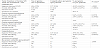
We included 333 eligible patients. Table 1 shows the characteristics of the study group. Among the 333 patients, 525 groins were dissected, 192 patients (57.7%) undergoing a bilateral procedure and 141 (42.3%) a unilateral procedure. Inguino-femoral lymphadenectomy was performed in 278 patients (79.7%) (416 groins), a nodal debulking in 65 patients (18.6%) (103 groins), and a sentinel node biopsy in 6 patients (1.7%) (6 groins). The median number of nodes removed per groin was 9 for patients having an inguino-femoral lymphadenectomy, 3 for a nodal debulking and 2.3 for a sentinel node procedure.
Sixty-nine patients (20.7%) received adjuvant radiotherapy to the groins and pelvis, while 12 patients (3.6%) received primary radiotherapy to the vulva and both groins. All 12 patients underwent some form of groin node procedure prior to their radiotherapy.
Groin wound drains were used in 211 patients (63.4%) and 348 groins (66.3%), with the drain left in-situ for a median of 6 days (range 2 - 16). Overall median length of post-operative hospital stay was 13 days (range 2 - 65) and was significantly longer when a groin drain was used (14 days versus 10 days respectively, p = 0.005). The median follow-up was 49 months (range 6 - 366 months). Twenty-two patients (6.6%) were excluded from the analysis for long term complications (lymphedema and recurrent lower limb cellulitis) due to follow up of less than 6 months. Eleven of these patients died within five months of surgery (4 of progressive disease), and 11 were lost to follow up.
3.1 Short-term complications of the groin dissection
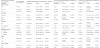
The commonest immediate post-operative complication was lymphocyst formation which occurred in 36.6% of the groins dissected (Table 2). There was no difference in lymphocyst incidence in groins having an inguino-femoral lymphadenectomy before 1991when the fascia lata was resected compared to after 1991 when the fascia lata was preserved (39.4% vs 42.7% respectively, P = 0.4). Lymphocyst formation was most strongly associated with a greater number of nodes removed (p = 0.0001) (Table 3). When adjusted for other risk factors, the number of nodes removed remained statistically significant for lymphocyst formation (p = 0.0001; OR 1.24 [95% CI 1.12-1.36] per node) (Table 4).
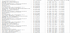
Univariate analysis indicated no difference in the incidence of lymphocyst formation when a groin drain was used. There was a bias in the indication for the use of drains, as they were more commonly used following an inguino-femoral lymphadenectomy (72.4%) than following nodal debulking, (43.8%) (p < 0.001). Use of a drain compared to no drain resulted in no significant difference in the incidence of lymphocyst formation for either an inguino-femoral lymphadenectomy (39.7% vs 48.7% respectively, p = 0.121) or nodal debulking (17.4% vs 13.5% respectively, p = 0.647) on univariate analysis. After adjusting for the number of nodes removed, patients having more nodes removed had a lower rate of lymphocyst formation with a groin drain, but this failed to reach statistical significance (p = 0.06) (Table 4).
The next most common short-term complication was groin wound infection, which occurred in 10.7% of the groins dissected. This was more common in the groins having an inguino-femoral lymphadenectomy (11.3%) than a nodal debulking (7.8%), but the difference was not significant in univariate analysis (p= 0.4) (Table 2). However, in multivariable analysis, increasing number of nodes removed was associated with an increased incidence of groin wound infection (p = 0.02) (Table 4).
The least common short-term complication was groin wound breakdown, which occurred in 8.2% of groins dissected (Table 2). In univariate analysis, the factors significantly associated with a higher rate of groin wound breakdown were increasing number of nodes removed (p = 0.005), current smoking (p = 0.02) and obesity (p < 0.001) (Table 3). On multivariable analysis, increasing age was also associated with groin wound breakdown (p = 0.02; OR 1.74, [95% CI 1.11 – 2.74] per 10 years), along with current smoking (p = 0.02) and number of nodes removed (p = 0.04) (Table 4).
3.2 Long-term complications of groin node dissection
Lymphedema was the major long-term complication occurring in 31.6% of the groins dissected. Lymphedema was more common in groins having an inguino-femoral lymphadenectomy (35%) compared to those having a nodal debulking (19.6%) or a sentinel node procedure (0%) (p = 0.003) (Table 2). An increasing number of nodes removed was significantly associated with an increasing incidence of lymphedema (p = 0.003) (Table 5).
On univariate analysis, there was evidence that obesity was associated with an increased incidence of lymphedema (p =0.01)(Table 5). However, no significant association was found in the multivariable analysis, where BMI was included as a continuous variable (Table 4).
When radiotherapy to the groin was included in our multivariable analysis, the wide 95% confidence interval did not allow a strong conclusion to be drawn about its association with lymphedema (p = 0.4; OR 1.61 [95% CI 0.53 - 4.90]) Table 4.
Recurrent lower limb cellulitis was documented in 6.8% of patients (Table 2), but this was probably substantially under-reported because over 50% of our patient population were referred from regional and rural areas and would have been treated for this complication locally. For this reason, recurrent cellulitis was excluded from further analysis.
4. Discussion
This is one of the largest series in the literature reporting on groin morbidity following groin node dissection for vulvar cancer. The principal findings were the relatively high incidence of lymphocyst formation and lymphedema, and the relatively low incidence of groin wound breakdown and infection, despite routine resection of the saphenous vein.
Our lymphocyst incidence of 36.6% per groin falls within the reported range of 13% to 60% [10-15]. The incidence of lymphocysts increased significantly as the number of nodes resected increased.
The issue of drains is controversial. In view of the relatively high incidence of lymphocysts despite drain usage, the senior author began to omit the insertion of a drain in 2002. Instead, Camper’s fascia was firmly sutured to the underlying fascia lata. Lymphocysts continued to be a problem but another recent Australian study has also reported no statistically significant difference in lymphocyst formation between patients with and without groin drains [15]. In a prospective Dutch study where drains were routinely used, the incidence of lymphocyst formation was reported to be lower when the drain was left in situ until drainage was < 30mls (range 2-40 days), compared to routine removal on the 5th post-operative day (16 % versus 60% respectively) [14].
The post-operative drain management after axillary lymphadenectomy for breast cancer has been studied more extensively. Two recent studies of breast cancer patients have also concluded that the use of an axillary drain did not significantly affect symptomatic seroma rates, or any other wound complication rates [16,17]. In both these studies, post-operative hospital stay was significantly longer in the drainage groups, which was also our experience.
Our wound infection rate of 10.7% per groin is low when compared to other studies, where incidences are reported to range from21% to 59% [10,12,14,15,18]. The only risk factor we identified was an increasing number of nodes removed. One recent study found that the incidence of post-operative groin cellulitis was lower in patients without a groin drain [15].
Our 8.2% incidence of groin wound breakdown is one of the lowest rates reported [10,11,13,14,18-20]. In addition to increasing number of nodes removed, increasing age was also a significant risk factor. This association has been noted in some studies [11,20], but not in others [10,12].
A Gynecologic Oncology Group study reported that the presence of a drain significantly increased the risk of groin wound breakdown [21]. Our groin breakdown rate was also higher in patients having a drain (9.8% versus 5.1%), but this was not significant on either univariate or multivariable analysis.
As expected, we found that current smokers were at a higher risk for groin wound breakdown. To our knowledge, only one other vulvar cancer study has reported this association [12]. However, two recent studies from the United States have reported significantly increased wound dehiscence rates in smoking cohorts. One study involved plastic and general surgical patients [22], and the other patients undergoing radical cystectomy [23].
The issue of saphenous vein preservation versus resection is controversial [19,20,24]. Two reports have suggested that saphenous vein preservation decreases groin wound breakdown, but the incidence of groin breakdown in these papers (13% and 16% respectively) was higher than our 8% incidence with vein resection [19,20]. Other studies have reported no correlation between saphenous vein ligation and complication rates for the groin dissection [10,13]. We believe that the most important aspect of preventing groin wound breakdown is the preservation of all the subcutaneous fat above Camper’s fascia.
The reported incidence of lymphedema ranges from 10.9% [25] to 67% [21]. Our overall incidence of 31.6% per groin is in the midrange of those reported [13,15,19,20,24,26,27]. The incidence was strongly correlated with the number of nodes resected and most studies concur with this finding [12,18,20,25,28]. Some authors have suggested that preservation of the saphenous vein may decrease the incidence of lymphedema [19,20], but as the problem is related to lymphatic obstruction, not venous congestion, this hypothesis lacks biologic credibility.
We have previously reported that nodal debulking for patients with bulky positive groin nodes followed by post-operative groin and pelvic radiation does not compromise survival [29], and the procedure is applicable to all patients with bulky positive nodes. The safety of the procedure was recently confirmed in a study from Leiden University [30]. In our experience, only 3.9% of groins experienced a wound breakdown after a lymph node debulking, 7.8% a wound infection and 14.6% developed a lymphocyst. These data support the earlier initiation of post-operative groin and pelvic radiation and would suggest that nodal debulking rather than an inguino-femoral lymphadenectomy should be considered the treatment of choice for patients with bulky positive nodes.
In accordance with two previous studies [10,25,28], and in contrast to two other studies [20,28] we found no significant association between the incidence of lymphedema and the addition of groinradiotherapy, although the relatively small numbers having radiation therapy may have not provided sufficient power to detect important differences.
Like some earlier studies [10,11], we found evidence of an association between the development of a lymphocyst and the subsequent development of lymphedema in univariate analysis (p = 0.04), although the evidence was weaker after accounting for other factors (p = 0.06). Obesity was also found to be a risk factor on univariate but not multivariable analysis, possibly due to the small number of patients in the higher BMI range. To our knowledge, higher BMI as a risk factor for developing lymphedema has only been reported in two other studies [28,31].
The major limitation of this study is the retrospective nature of the review. The incidence of long-term complications, particularly recurrent cellulitis, may have been under-reported because over 50% of the patients came from rural areas, and some were only seen annually. The strengths of the study are its large sample size, its per groin analysis, and the management of all patients in one specialised unit with a common treatment protocol.
5. Conclusions
Appropriate groin node dissection is a critical part of the treatment for all patients with vulvar cancer, except those with stage IA disease. Lymphocyst formation in the immediate post-operative period and lymphedema after several months are the major morbidities, and both are associated with the number of lymph nodes removed. In this study, the use of groin drains did not significantly decrease the incidence of lymphocyst formation. Groin node debulking for all patients with bulky positive nodes, and sentinel node biopsy for patients with small primary tumours are the only legitimate ways to reduce the number of resected groin nodes. Groin wound breakdown should occur in less than 10% of groins if care is taken to preserve the subcutaneous fat above Camper’s fascia, regardless of resection of the saphenous vein.
Competing Interests
The authors declare that they have no competing interests.
Author Contributions
The authors declare their responsibility for the content of this publication. EB and NH developed the concept of the article. EB conducted the data collection and analysis and drafted the manuscript. NH edited all drafts. MD provided statistical guidance and assistance with interpretation and reporting of results. All authors approved the final version for submission.