


1. Introduction
Endometriosis is defined as the presence of endometrial stroma and glands outside the normal uterus. As reported in the past by the Endometriosis Association Registry a total of 38% of women diagnosed with endometriosis may have symptoms before the age of 15, while a mean number of 4.2 physicians have examined the adolescent before final diagnosis is set [1]. On the other hand, a range between 19% and 73% of adolescents undergoing laparoscopy for chronic pelvic pain are diagnosed with endometriosis. The same was found by a study of Goldstein et al. [2] who reported that the prevalence of endometriosis found at laparoscopy in a prospective study of adolescent females with pelvic pain is 47%, while other studies have shown a prevalence of 25- 38% for these adolescents. [3,4]. A 66% of adult women have reported the onset of pelvic symptoms before the age of 20 according to the Endometriosis Association.
It is of great importance that 50-70% of adolescents with pelvic pain, who have received Combined Oral Contraceptives (COCs) and/or Nonsteroidal Anti-Inflammatory Drugs (NSAIDs), but no responding to them, have signs of endometriosis during laparoscopy. Interestingly, endometriosis has also been identified in premenarcheal girls with some breast development [5,6]. This can be explained by the theory of embryonic müllerian rests or coelomic metaplasia as opposed to retrograde menses.
2. Pathophysiology
Several factors have been incriminated for endometriosis, while no single theory can explain the variety of symptoms. Sampson [7] was the first who reported that during menstruation endometrial cells regurgitate through the fallopian tubes and implant in the pelvis. Another theory has proposed that metaplastic cells transform into endometrial cells, [8] and these metastasize through lymphatic and vascular channels, resulting in endometriosis [9]. This theory can explain the findings of endometriosis in other tissues such as the lung, brain, and skin. Other multi-factorial hypotheses with immunological, anatomical and genetic mechanisms, leading to dysfunction in the ectopic endometrium can explain several other endometriosis cases. Theoretically, all women should have been diagnosed with endometriosis, due to normal retrograde menstruation at the pelvis in every cycle [10]. The above theories, explain why some women diagnosed with endometriosis and others not, paying attention in individual features, such as family history of endometriosis, early menarche and exposure to circulating steroid hormones, body mass index during late childhood and early adolescence. Moreover, lifestyle characteristics and environmental factors are likely related to the development of the disease playing an epigenetic role. Positive family history has been reported by many studies [11], even though this association cannot only be explained by genetic mechanisms. It is important that the disease among first-degree relatives is six to nine times higher than in the population [12,13].
An early menarche is also positively associated with endometriosis [14], due to the fact that these girls are more likely overweight, with higher levels of adipose fat tissue and circulating steroid hormones [15,16]. Another factor that seems to play a role in the inverse relation between childhood and early adolescence body size and the incidence of laparoscopically confirmed endometriosis is anovulation due to insulin resistance and hyperinsulinemia in obese pre-adolescent girls [17].
Finally, endometriosis can only be diagnosed by visual inspection during laparoscopy, ideally confirmed by histology and can present as peritoneal disease with typical or subtle lesions, ovarian endometriotic cysts or deeply infiltrative disease or as a combination of these features. Different Classification systems have been used at the past in order to set different stages of the disease. The degree of endometriosis can be staged by laparoscopy as minimal, mild, moderate or severe according to the classification of the American Society of Reproductive Medicine [ASRM-former American Fertility Society (AFS)] [18]. Studies regarding the ASRM classification system have shown that adolescents with endometriosis, hadeither minimal (50%), mild (27%), moderate (18%) or severe (14%) disease. Other classification systems include the Endoscopic Endometriosis Classification I-IV by Semm (EEC I-IV) [19], the Acosta classification [20] or the staging system proposed by Kistner et al. [21] with a scale of I-IV. The variety of studies and classification systems agree that the prevalence and severity of the endometriosis is believed to significantly increase with age therefore is considered as a progressive disease [22].
3. Diagnosis
The main symptoms during diagnosis of endometriosis in adolescence, is chronic pelvic pain (27%-96%) and dysmenorrhea (18%-100%) [23,24]. Acyclic pain seems to be more common in adolescents than in adults. Other symptoms that can support diagnosis are gastrointestinal symptoms, urinary symptoms, irregular menses, dyspareunia, pelvic mass, subfertility, constitutional symptoms and depression/anxiety [23]. In all adolescents is offered apain diary, in order to document frequency and all characters of pain. Smorgick et al. have reported that the prevalence of comorbid chronic pain syndromes (56%) and mood disorders (48%) in adolescents suffering from endometriosis is not uncommon, while irritable bowel syndrome, interstitial cystitis/painful bladder syndrome and chronic headaches can be found in up to 25%, 16% and 19% respectively in these adolescents [24].
In a study by Laufer et al. [25], 90.6% of adolescents with endometriosis had acyclic pain versus 69% in the adult population as reported above [26]. Müllerian anomalies, especially those with outflow tract obstructions, are statistically significantly positively correlated with endometriosis, being an independent risk factor. This was shown by a study be Yang et al [27] who reported that genital tract malformations can present in up to 24%of patients with endometriosis. The majority of adolescents have early stage disease, but up to 33% of them have advanced disease. Fedele et al. [28] found no correlation between severity of pain symptoms and stage of the disease or site of the endometriotic lesions, while an ovarian endometrioma is the most common presentation of advanced endometriosis in adolescents. Recent studies have report a large number of cases of adolescents with Stage III and IV endometriosis. The adult literature reports Stage I disease in 30%-39%, Stage II in ~12%-13%, Stage III in 27%-35% and Stage IV in 13%-28% [29,30]. Even though adolescents may present with advanced stages of endometriosis, these number are fewer comparing with adults.
Red lesions are the most common lesions seen in adolescents, with atypical lesions being common as well. Two studies, one by Davis et al [31] and another by Reese et al [32] showed that the vast majority of lesions in adolescent populations with endometriosis are red lesions, while a large number of these lesions were correlated with severe dysmenorrhea, with complaints of abdominal pain, nausea, constipation and diarrhea. Another study reported atypical red vascular lesions in 60% of adolescents compared to only 20% of nonadolescents [30]. Clear lesions are common in adolescent endometriosis but often difficult to visualize and evaluate. Peritoneal defects, or windows, which are possible manifestations of endometriosis, are very common in adolescents. The reported incidence in adolescents is around 10%-18.4% as quoted in several studies [33].
Past medical history, family history and physical examination are mandatory during evaluation and management of adolescents with a possible endometriosis. Several other pathologies, such as appendicitis, pelvic inflammatory disease, müllerian anomalies or outflow obstruction, bowel disease, hernias, musculoskeletal disorders, and psychosocial complaints should be excluded in order to set the diagnosis. Inspection of the girl for a possible estrogendominant body configuration with peripheral fat distribution and for breast and pubic hair development according to the Tanner classification system is of great importance [23].
A patent outflow should be performed in all adolescent by placing a Q-tip into the vaginal canal. This is very helpful, in order to exclude a possible transverse vaginal septum, vaginal agenesis, or agenesis of the lower vagina. For virgo adolescents pelvic examination cannot be performed, therefore, a rectal-abdominal examination, even discomfort for adolescents, in the dorsal lithotomy position, may be helpful to determine if a pelvic mass is present. Attention should be given in the existence of both diffuse and focal pelvic tenderness [23].
Imaging exams are very helpful during evaluation of these girls. Ultrasonography and magnetic resonance evaluate anatomical structures, but are not specific for diagnosing of endometriosis. According to some studies, MRI can detect endometrial implants with a sensitivity as high as 60%, while this method can be used in order to follow up adolescents’ response to treatment, even though its cost is high [34]. Blood tests, such as CA 125, are very sensitive, but it is not specific and, thus, is not helpful in the diagnosis of adolescent endometriosis. No data exist regarding the use of CA 125 to monitor the clinical progression or regression of disease in adolescents with endometriosis [35].
Symptomatic adolescents should be evaluated laparoscopically when standard treatment of pelvic pain or dysmenorrhea is not effective. Endometriosis should be staged using the revised criteria of the American Society of Reproductive Medicine point-based classification system as mentioned above [18]. Biopsying during laparoscopy sites of apparent endometriosis, especially atypical lesions, in order to confirm the diagnosis and avoid mislabeling a patient is of great importance, while biopsying normal appearing peritoneum should be left at the surgeon's discretion because it is somewhat controversial [36].
Progression of the disease in adolescents has been a topic of argument among researchers. In 2010, Unger and Laufer [37] published the case reports of three adolescents, aged between 13 and 16 years, suffering from severe pelvic pain and diagnosed with Stage I endometriosis at the time of laparoscopy. Reese et al. [32] presented 39 adolescents, with 4 patients (18%), in the two older age groups (16- 17 and 18-20 years), suffering from Stage III or IV. Tandoi et al. [38] studied 57 women aged 21 years or younger over a 5-year period and in 32 (56%) observed a recurrence of the disease after surgery. Its rate increased with time from surgery, with no apparent association with site or stage of the disease, type of surgery, and post-surgical medical treatment. Yang et al. [27] reported that 45.7% of 35 adolescents included in the study, suffered from disease recurrence with an average time of recurrence of 33.4 months.
4. Treatment
Treatment algorithm for adolescents presenting with dysmenorrhea according to the American College of Obstetricians and Gynecologists is summarized in Figure 1 [39].
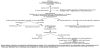
Recommendations include initiation of treatment with NSAIDs and COCs. In case of symptoms persistence, after 3 months, a diagnostic laparoscopy should be offered for these girls [39]. As reported above, lesions seen in adolescents’ pelvis, during laparoscopy are different from the typical powder burn lesions seen in adults [40]. Endometriosis symptoms control, prevention of disease progression and preservation of fertility are primary goals of treatment. Medical and surgical options are available for the management of endometriosis.
4.1 Medical management
4.1.1 NSAIDs
NSAIDs can be used as empiric treatment during management of dysmenorrhea in adolescents, even though the diagnosis of endometriosis has not been set yet.
4.1.2 Combined oral contraceptives (COCs)
COCs are typically the first line treatment and can be used as well as empiric treatment. Acting by ovulation inhibition, they decrease gonadotropin levels and therefore reduce menstrual flow and cause decidualization of endometriotic implants. Another role of COCs is the decrease of cell proliferation, as well as the reduction of eutopic endometrium. COCs can be used as continuous treatment in order to induce amenorrhea, with therapy being suppressive and not curative, while stopping treatment for more than 6 months, can lead to symptoms recurrence. Finally, according to Cochrane Database of 2007 there are no sufficient data regarding long-term benefits of COC in the treatment of endometriosis [41].
4.1.3 Progestins
Progesterone agents include medroxyprogesterone acetate (MPA) and 19 nortestosterone derivatives, such as norethindrone and norgestrel. These agents lead to decidualization and atrophy not only in ectopic, but as well in eutopic endometrial tissue. 20 to 30 mg daily or the depot form of 150 mg every 3 months of medroxyprogesterone acetate can be used in order to treat symptoms of dysmenorrhea or adolescents’ endometriosis. It is important that up to 70% to 80% of girls suffering from endometriosis show symptoms improvement. On the other hand the benefits of long-term use of progestin therapy needs to be weighed against impaired bone mineralization secondary to the hypoestrogenic environment induced by progestins, with the risk for osteoporotic fractures been yet unknown, as reported also by the U.S. Food and Drug Administration [42,43]. Other side effects include weight gain, bloating, mood lability and irregular bleeding.
4.1.4 Danazol
Danazol is a 17-ethinyl testosterone derivative with an efficacy being equivalent to a variety of GnRH agonists in treating endometriosis. Its androgenic effects, affecting sex-hormone-binding globulin levels, resulting in an increase of free testosterone. Buttram [44] had studied 220 patients, complaining about weight gain, depression, muscle cramps, decreased breast size, flushing, oily skin and hair, acne, hirsutism, irreversible deepening of the voice, and skin rash. Among them 7% discontinued the drug secondary to intolerable side effects and this is enhanced by the fact that patients using GnRH agonists reported a better quality of life compared to them using danazol. Finally, due to the fact that this agent is poorly tolerated by adolescents is not widely utilized in endometriosis management [45].
4.1.5 Cyproterone acetate
Cyproterone acetate (CPA) is a 17-hydroxyprogesterone derivative with antiandrogenic and antigonadotropic properties. As reported in a study by Fedele et al. [46] 27 mg/day oral CPA with 0.035 mg ethinyl estradiol could be used to treat women with endometriosis successfully, while another study by Vercellini et al. [47] including 90 women, who received either 12.5 mg/day CPA or a daily COC (0.02 mg ethinyl estradiol + 0.15 mg desogestrel) for 6 months, showed that after 6 months pain scores were reduced in both groups. This supports the idea that girls in whom estrogens are contraindicated, CPA may be an alternative sufficient treatment.
4.1.6 GnRH agonists
GnRH agonists are very effecting in treating adolescent endometriosis and alleviate symptoms associated with endometriosis. Acting by inducing menopause with binding to the GnRH receptors in the pituitary, they result to cessation of pituitary gonadotropin release and subsequently to amenorrhea. According to the Cochrane Group reviewed the efficacy of GnRH agonists versus COCs in the treatment of endometriosis, GnRH agonists are more effective than COCs. GnRH agonists include leuprolide acetate, nafarelin, buserelin, and goserelin. Leuprolide can be given as a 3.75-mg injection every 4 weeks or 11.25-mg injection every 12 weeks. It is of great importance to remember that the use of GnRH agonists alone is generally limited to patients more than 16 years of age and for a period no more than 6 months [48].
4.1.7 Add back therapy
In order to prevent side effects of pseudomenopause associated with GnRH agonist like vasomotor symptoms, vaginal dryness, and mood swings, hormonal ‘‘add-back’’ options are recommended. These include norethindrone acetate (5-mg daily) and combined conjugated estrogens/medroxyprogesterone acetate (0.625/2.5- mg daily). Adolescents accruing bone mass up to the age of 20 years, therefore initiation of GnRH agonists in this age should always begin in combination with add-back therapy, while BMD monitoring should be offered every 2 years. Calcium and Vitamin D supplementation should be given in all these girls in order to avoid bone demineralization [49].
4.2 Surgical treatment
Surgical options in adolescent endometriosis include laparoscopy rather than laparotomy. The role is both diagnostic and therapeutic and usually a specialized physician in laparoscopy and adolescent endometriosis is preferred to perform the procedure. Surgery should be timed in the follicular phase of menstrual cycle, in order to avoid future possibility of recurrences and adhesions. First port should be intraumbilical and the lateral ports should be placed close to the pubic bone for cosmetic superiority. The goal of surgical treatment is to remove visible areas of endometriosis and restore normal anatomy by lysis ofadhesions. The procedure seems to improving endometriotic symptoms in 38% to 100% of adolescents [50]. Laser vaporization, unipolar or bipolar coagulation, and endocoagulation are the methods used, with no one technique has been shown to be superior to any other.
Surgical treatment seems to improve pain in adolescents with endometriosis. This was summarized in a meta-analysis by Janssen et al. [51], in which girls were treated either with ablation or excision of endometriosis and pain improvement was shown. Furthermore, Yang et al. [27] concluded that there was a decrease in chronic pelvic pain (by 23.5%) and dyspareunia (by 11.8%) after complete excision of endometriotic lesions, while in a study be Dun et al. [23], 64% reported resolved pain and 16% reported improvement of pain at 1 year after the laparoscopic excision and ablation of lesions.
Surgical treatment can also improve fertility options in adolescence. In a retrospective case series to assess the long-term fertility outcomes in young women after laparoscopic surgery (excision and ablation) for endometriosis, a long-term pregnancy rate of 71.4% of which >80% were achieved without assisted reproductive technology (ART) was shown, with most of the patients who conceived had Stage I/II disease [52].
Surgery is also very helpful in reducing disease progression and/ or recurrence. In some studies is reported that complete laparoscopic excision by experts can significantly reduce the recurrence rates of endometriosis in adolescents, while Yang et al. [27] found zero rate of recurrence (diagnosed visually or histologically) after complete laparoscopic excision of the disease in teenagers at a repeat laparoscopy for pain. Even though the frequency of adolescents undergoing laparoscopy for persistent recurrent pain is 47.1% the rate of endometriosis found at surgery was zero [53].
On the other hand, postoperative hormonal suppression should be offered to adolescents in order to treat symptoms and to prevent progression and/or recurrence of the disease, while the role of postoperative medical therapy in conjunction with surgery in improving future fertility of adolescents with endometriosis has not been evaluated. Moreover, the conjunction of surgery with postoperative medical therapy does not seem to slow disease progression and/or recurrence. The recurrence rate of endometriosis in young women appears to be higher than in older women. In a retrospective cohort study of 57 women, aged ≤21 years, who were treated initially by excisional surgery, was shown that the rate of recurrence of symptoms during a follow up-period of 5 years was 56%. The study also showed that the postoperative medical therapy did not influence the recurrence rates [38].
As reported above, a variety of medical therapies have been used in treating endometriosis during adolescence. Even though further studies needed, in order to conclude which medical therapy is superior to another, GnRH agonists seem to be more effective compared with COCs and progestins to prevent disease recurrence. Combination norethindrone acetate plus conjugated equine estrogens as add-back therapy, appears to be more effective for increasing total bone mineral content, bone mineral density and lean mass than norethindrone acetate monotherapy [54,55]. Levonorgestrel intrauterine system (LNG-IUS) is accepted for use in the adolescent population for contraception and menorrhagia, but there are not enough data regarding its effectiveness in the treatment of adolescent endometriosis.
4.2.1 Endometrial cysts
There are very few data about endometriosis and endometriomas in adolescents and young women. A study reviewing 15 years of ovarian masses in infants, children and adolescents reported no endometriomas [56]. Ovarian endometriomas are usually correlated with more advance stage of the disease.
As it is easily understood, these girls are present with more frequent pain. A retrospective study of 63 adolescents with endometrioma found bilateral disease in 22.22%, while endometrioma in the right ovary seems to be more frequent than a left endometrioma (65% vs. 57%). In these cases the preferable surgery is a combined technique of cystectomy and cauterization of the capsule [57]. Interestingly, in a review by Gordts et al. [58] was found that early ablative surgery can contribute to a lower morbidity, relief of symptoms, and a better quality of life, while in another recent published study have reported recurrence rates of endometrioma per patient at 24, 36, 60 and 96 months after laparoscopic cyst enucleation for ovarian endometrioma being 6.4%, 10%, 19.9% and 30.9%, respectively. All these adolescents had stage III or IV of the disease.
5. Future Trends
Selective estrogen receptor modulators (SERMS) and selective progesterone receptor modulators (SPRMs) are the new treatment options for adolescent endometriosis. However, we have not yet human studies for both drug groups. Both are acting by suppressing estrogen dependent endometrial growth without the adverse systemic effects of hypoestrogenism, like vasomotor symptoms and loss of BMD. Another treatment options, is the use of aromatase inhibitors. This is a key enzyme in estrogen biosynthesis, appears to be over-expressed in sites of endometriosis. This drug acting by reducing ovarian and local production of estrogens, therefore can be used in treatment of adolescent endometriosis. Finally, autoimmune modulators may be an effective method of disease treatment, with anti-tumor necrosis factor therapies have been already successfully used to reduce endometriotic growth in animal models, being a promising future treatment method [59,60].
5.1 Follow-up
A careful follow-up of adolescents with endometriosis is mandatory, due to the fact that it can be a life-long disease. Patient should be examined every 3 to 6 months. During this time, a pain calendar should be used in order to monitor the pain, while concerns regarding future fertility and quality of life should be addressed. Unless contraindicated, most patients should be put on COCs after surgery. If the patient does not respond to surgery or has recurrence of symptoms, other treatment modalities should be considered. A multidisciplinary approach is usually needed, including a gastroenterologist, psychologist and urologist.
5.2 Issues for Future Consideration
Future studies should be focused on the role of early diagnosis of endometriosis and treatment in progression and advancement of the disease. Question remaining regarding, how the adolescent daughter of a woman, who had no pelvic pain, but stage IV endometriosis and infertility should be treated. Is it important for that girl to be evaluated and treated, even though she has no pelvic pain, in order to exclude the possibility of endometriosis.
6. Conclusions
Adolescent endometriosis is not uncommon being a progressive disease. Pain is the main symptom, especially dysmenorrhea and chronic pelvic pain. NSAIDs and COCs can be used as first line medical treatment during management of adolescent endometriosis, but in case of symptoms persistence, the girl should undergo a laparoscopy, with the likelihood of endometriosis during laparoscopy being up to 50%. The appearance of lesions found at laparoscopy in adolescents may differ from that in adults, while COCs, DMPA, GnRH agonists and surgical ablation tend to be the practical treatment modalities in this age group. Primary goals are alleviation of pain symptoms, avoidance of disease recurrence and assurance of future fertility preservation. A multidisciplinary approach to pelvic pain with the assistance of pain treatment services is usually recommended, while future work for adolescents should focus on developing safe, minimally invasive, treatment techniques.
Competing Interests
The authors declare that they have no competing interests.