


1. Introduction
Choriocarcinoma was the fourth gynaecological cancer cause of death at the Korle Bu teaching Hospital, Ghana, after cancer of the cervix, ovary and endometrium [1] . In that study, the commonest gynaecological cancers in women under 30 years of age were ovarian cancer (52.2%) and choriocarcinoma (41.5%) [1] . All other studies found a mean age of 29 years and peak age of 25 – 29years [2] . The crude prevalence rate in Korle-bu Teaching Hospital is 0.34/1000 deliveries while it's 0.33/1000 in Uganda [2] . Choriocarcinoma can be preceded by; hydatidiform mole, spontaneous abortion, ectopic pregnancy and rarely a normal term pregnancy [3,4]. Choriocarcinoma preceded by live birth is reported to occur in about 1 in 50,000 live births in the United Kingdom and is reported to be associated with a poor prognosis [3] . A number of such cases have been reported in the literature [3,4] .
Gestational choriocarcinoma can present as abnormal bleeding per vaginum or with evidence of tumour metastasis to other organs such as the lung or brain resulting in cough with bloody sputum production or a stroke [4] . The primary lesion may be extensively necrotic and be absent with the secondary lesions being the only source of clinical manifestation [4] . Laboratory titers of beta human chorionic gonadotropin are elevated to levels above those encountered in hydatidiform moles. Occasional tumours, however, produce little hormone, and some tumours become so necrotic that they become functionally inactive [5] .
Detailed clinical, postmortem gross and histopathological analysis of a patient who presented with antepartum haemorrhage and subsequently severe persistent postpartum haemorrhage even after a caesarian hysterectomy and finally a stroke is presented and histopathological findings correlated with clinical findings.
2. Case Report
A 27 year old mother of three was rushed to the obstetric emergency room on account of heavy bleeding per vaginum and symptoms of anaemia one (1) month after delivery through a Caesarean hysterectomy. She had a history of two previous Caesarean sections. Her baby from her recent delivery was reported as being well. The indication for her recent Caesarean delivery was a history of two (2) previous Caesarean sections and an impression of ante partum haemorrhage due to a suspected placental praevia, a Caesarean hysterectomy was however done for her due to a morbidly adherent placenta. There were no imaging studies at the time of that admission in line with the local practice that women with a history of two previous Caesarian sections are delivered through another Caesarian section. Antepartum haemorrhage further reinforced the need for the Caesarian section, only this time as an emergency. The Caesarian hysterectomy specimen was not sent for histopathological examination. While on admission prior to surgery, she had episodes of haemoptysis and was referred to the Ear Nose and Throat (ENT) surgeon but no documentation of the ENT review was seen in the patient’s clinical notes. At her last presentation, she was afebrile but pale. She was not in respiratory distress and her chest was clinically clear. Her pulse rate was 90 beats per minute and Blood Pressure (BP) 100/60mmHg. Her abdomen had a Pfannenstiel scar, was soft, non-tender, with no palpable masses. Vaginal examination revealed a large necrotic mass at the vault of the vagina that bled with contact. A sample was sent for histopathological analysis. A diagnosis of Secondary post-partum haemorrhage due to retained placental tissue, to rule out choriocarcinoma was made. She was admitted and transfused a unit of blood and started on antibiotics and haematinics. Laboratory investigations showed; Haemoglobin 6.7g/dl (12-18), White cell count 8.5x10³ (2.6-8.5 x 10³), Platelets 233 x 103 (140-400 x 103) and Beta HCG 2533.2 mIU/ml. Nine days after admission the patient developed sudden onset of severe headache, was said to be agitated with slurred speech, right facial palsy and left hemiplegia. Her chest had scattered bilateral basal crepitations. Her BP was 100/40mmHg. Her neck was supple and Kernig’s sign was negative. A diagnosis of; Cerebrovascular Accident with Left hemiplegia due to a Metastatic choriocarcinoma, was made to be confirmed with a Computer Tomography scan of the head but the patient died shortly. Chemotherapy had not been initiated.
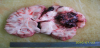
At autopsy, the external examination was unremarkable except for pallor of mucous membranes, a healed pfannenstiel incision and a blood stained vulva. Internal examination showed multiple nodules, some haemorrhagic, ranging in size from 3mm to 2cm across, distributed in the parenchyma of all lung lobes and on the pleural surfaces of both lungs (figure1). No ovary, uterus or cervix was seen. Instead, a haemorrhagic mass measuring 10cm across was seen in the pelvis. The vaginal vault had a similar haemorrhagic and necrotic lesion continuous with the haemorrhagic pelvic mass. There was a right intracerebral cerebral haemorrhagic lesion, 8cm across with liquefactive necrosis of the surrounding brain tissue with haemorrhage extending into the subarachnoid space (figure 2). The brain was edematous. Histology of tissue from the pelvic mass, vaginal vault, lungs and brain showed similar features; a tumour with large areas of haemorrhage and necrosis composed of an admixture of bizarre syncytiotrophoblasts and cytotrophoblasts. No chorionic villi were seen (figure3), in keeping with choriocarcinoma.


3. Discussion
Post-partum choriocarcinoma is a very rare complication following normal live birth [3] . The index patient presented with antepartum haemorrhage and postpartum haemorrhage and was initially thought to have a low lying placenta before delivery and then retained products of conception post-partum. These diagnoses are amongst the commonest causes of antepartum and postpartum haemorrhage in poor resource settings and compounded by cost implications, may have informed the decision of the clinical team to manage as such. It likely informed the decision not to present the Caesarian hysterectomy specimen for histopathological analysis and also, the absence of imaging studies. It is however reported that a pelvic ultrasound is often useful to detect the extent of uterine involvement and may identify patients who are at risk for uterine perforation or who would benefit from a hysterectomy to reduce tumor burden. Furthermore, a chest X-ray helps to evaluate lung metastasis though if negative, a chest computed tomography (CT) scan may be performed since it may detect micrometastases in 40% of patients with a negative chest X-ray [6] . It is obvious in the index case that a chest X-ray could have aided immensely in the diagnosis and early management of this patient despite the poor prognosis [6] .
In a study by Dobson et al, it was reported from their major experience over 25 years that choriocarcinoma following a term non-molar pregnancy had poor prognosis. If staged, the index case had high risk disease (stage IV: >/= 7) evidenced by the presence of tumour in the lungs and brain, and the occurrence of tumour after a term pregnancy and its presence outside the uterus. These conferred a rather poor prognosis on the patient. The presence of tumour in the lungs and vagina meant the patient had a risk of cerebral metastases as reported in the literature [6] . Such high-risk choriocarcinoma is treated with multiagent chemotherapy [3] . The preferred agents used for the treatment of such high risk choriocarcinoma are EMACO (Etoposide, MTX, ACT-D, Cyclophosphamide, and Vincristine), it has the best effectiveness-to-toxicity ratio. It is reported to induce complete remission in 6 of 6 patients (100%) with high-risk stage II disease at one center, 36 of 37 patients (97.3%) with high-risk stage III disease at another and at other centers, it was reported that EMACO induced remission in 86% and 76% of patients with high-risk metastatic disease at two other centers [6] . Our patient did not benefit from any of these regimens. Combination chemotherapy is often administered at two- to three-week intervals and timely administration is essential [6] .
As in our case Dobson et al were concerned by the delay between onset of symptoms, diagnosis and subsequent referral for treatment. In the index case, considering that the patient presented with hemoptysis at the time of first presentation, when the caesarian hysterectomy was done (4 weeks to her demise), sending the caesarian hysterectomy specimen for histological analysis, performing a β hCG analysis, requesting a chest X-ray or having some index of suspicion for the persistent abnormal bleeding per vaginum resulting in the Caesarean hysterectomy would have led to earlier diagnosis and most likely averted the patient’s death. Delay in diagnosis is reported to increase patient morbidity from choriocarcinoma and may result in more frequent usage of higher dose multiagent chemotherapy [3] . Though the baby was reported as doing well, close follow up may be warranted considering that in the study by Dobson et al, it was reported that a baby delivered to one of their patients subsequently died of choriocarcinoma at 4 weeks [3] .
The index patient reported with signs of increased intracranial pressure and subsequently showed clinical signs of an intracerebral haemorrhage which was confirmed at autopsy to be due to metastasis. These findings were reported in a study by Suresh et al in India [7] . In that study, they emphasized the importance of considering metastatic choriocarcinoma as an important differential diagnosis of haemorrhagic intracerebral lesions in women of child bearing age [7] . They again emphasized the value of serum β hCG and histopathological examination of tissue for the diagnosis of such lesions. This is even more important in settings where choriocarcinoma is more common. The diagnosis notwithstanding, cerebral metastasis of choriocarcinoma confers a bad prognosis and in spite of the remarkable improvement in the treatment of choriocarcinoma, mortality is still significant; cranial metastasis being responsible for death in nearly 50% of cases [7] . It is thus essential that choriocarcinoma is diagnosed early and prompt treatment initiated to prevent death and reduce morbidity.
Our patient’s presentation is identical to that of a case reported by Lema et al [7] . Like our case, their case presented with antepartum haemorrhage and then secondary postpartum haemorrhage. At variance however is the morbidly adherent placenta that resulted in a caesarean hysterectomy in our case. In their report, they suggested that choriocarcinoma should be considered a possible local cause of antepartum and or postpartum haemorrhage in a parous woman, in areas where gestational trophoblastic disease (GTD) is relatively commoner, such as Africa [7] . This report underscores the need to rule out rare causes of antepartum and postpartum haemorrhage such as choriocarcinoma when managing patients presenting with such symptoms in our setting. Due to the unavailability of the caesarean hysterectomy specimen we are unable to comment on the histopathology of the placenta and what was clinically interpreted as a morbidly adherent placenta. This may well have been a focus of choriocarcinoma at its early stages. It again underscores the need for histopathological analysis of such specimen.
4. Conclusion
Choriocarcinoma is a rare cause of postpartum intracerebral haemorrhage, antepartum haemorrhage and postpartum haemorrhage and a high index of suspicion is required to diagnose the condition with readily available βhCG and histopathology. Once diagnosed however, current chemotherapy regimens assure a favourable prognosis. If not diagnosed early however, it is rapidly fatal.
5. Ethical Clearance
In our institution, ethical clearance is not required to report clinical cases once all identifiers have been removed from the report. Verbal consent was however sought from the relations.
6. Contribution
Frederick Hobenu conducted the autopsy and documented the findings photographically, Simon Naporo and Leonard Derkyi- Kwarteng reviewed the literature and Patrick Kafui Akakpo wrote the manuscript and also reviewed the manuscript. All the authors reviewed histopathology slides and the manuscript and agreed on its content.
Competing Interests
The authors declare that they have no competing interests.
Acknowledgments
We thank the histotechnicians in the department of pathology for preparing the slides for this case.