


1. Case Report
Since first description by Lobstein in 1816 until 1966 only 131 cases of lipomatous uterine tumours (UFT) were initially reported. From the 70’s up to date this number has been progressively growing to at least 357 cases around the world. The first preoperative study on a case of uterine “fatty” tumour has been described by Jacobs and Markowitz in 1988 [1] and since then the preoperative imaging study of these tumours has significantly increased due to the expansion of the radiological technologies and diffusion of ultrasonography in routine gynaecologic activity. Uterine fatty tumours (UFT) or lipoleiomyomas (LLM) are a kind of leiomyomas with prevalent fatty component occurring mostly in peri-menopausal and postmenopausal obese women. Many clinical and pathological aspects of these uncommon uterine nodules have been already reported [2-6]. Nonetheless literature largely varies with a prevalence of radiological case-reports for clinical data and histo-pathological review analysis on pathogenesis investigation. Little or nothing about UFT is found in important gynaecological journals. Moreover it is not clear which diagnostic tool among ultrasound, CT and MR should be best utilized for clinical follow up of these nodules [7,8], whether hysterectomy should or not always warranted [9,10] and how frequently a malignant transformation of these tumours should also be expected [3,11]. We report here a single case of UFT along with a full literature review on this topic with the aim to help gynaecologists in the clinical counselling of these tumours.
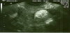
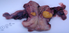
A 67 year old woman with no clinical symptoms or relevant medical history (menopause at 54 y.o., hypercholesterolemia and overweight [BMI=28.9]) was found with a 1.9 cm hyper echoic uterine nodule on the intramural/subserosal margin of anterior wall. This little hyper echoic nodule was clearly distinguished from the surrounding myometrium without showing any posterior acoustic shadow. Contrary to that generally reported this lesion appeared not to be encased in hypo echoic ring (Figure 1). The discovery of this nodule was made occasionally during routine transvaginal ultrasound (US). Following CT and MR (images here not reproduced) a presumptive diagnosis of uterine fatty tumour was made and patient advised to undergo surgery. A totally laparoscopic hysterectomy with bilateral adnexectomy was performed and successively the resulted histopathological analysis confirmed the benign nature of the lesion. Pathological findings: grossly, uterus weight of 76 grams and dimensions of 7 × 4 × 3 cm, at the cut surface with a yellowish intramural nodule of corpus uteri of 1.9 cm in diameter, with welldefined edges but non encapsulated; the overlying endometrium was atrophic and the cervix showed no significant macroscopic changes (Figure 2). Microscopically the tumour was predominantly made up of mature adipose tissue mingled with bundles of smooth muscle (positive for smooth muscle actin antibody at immunohistochemistry). The diagnosis of lipoleiomyoma was made. The patient gave her informed consent to the study.
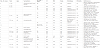
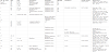
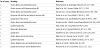
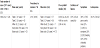
Overall a total of 226 fatty tumours of the uterus including lipomas, lipoleiomyomas, liposarcomas and endometrial cancers associated with lipoleiomyomas could be retrieved from the literature. We started reviewing data from the study of Willen et al 1978 [2] (the first 131 cases of UFT from 1816 to 1966 were excluded). This has corresponded to 75 independent studies here separately reported in 3 different tables: Table 1 for all clinical studies (n=46), Table 2 for pathological review data analysis (n=16) and Table 3 for malignant uterine tumours with fatty component and endometrial cancers coincidental with benign fatty tumours (n=13). Mean patient age, tumour size and incidence of UFT (which varied accordingly to the criteria used being low -0.03%- in hysterectomy and higher -0.8-2.1%- in uterine leiomyomas studies) along with symptoms and methodology used for preoperative tumour study and detection are summarized in table 4. No association whatsoever between aging and tumour size has been noted (Table 1). Despite being a tumour associated with aging we observed a prevalence of 23% in women younger than 54 years. As for all the other uterine fibroids, clinical signs largely varied including occasional discovery in asymptomatic patients, bleeding, abdominal or pelvic pain, pelvic mass discovery, liver problems, anaemia, disuria, and constipation. Despite UFT are said to recur more frequently in overweight and obese perimenopausal women, specific details on body weight or BMI have in general not been reported. Diagnosis and radiological follow up has been obtained in different ways including a variable utilization of multiple technologies such as US, CT, MR. All 3 of these have been employed only in 16/46 independent case reports. US either by transvaginal (TSV-US) or abdominal (TA-US) route was used in 42/46 studies but TVS has been applied only in 19 occasions. The use of TVS alone as unique diagnostic technique has been used only in 4 studies (for a total of 10 patients) while MR alone, as unique diagnostic tool, in 2 studies (for a total of 10 patients). Hysterectomy has been generally the rule for most patients but 9 (7 study reports). By a pathologist point of view, most of the uterine fatty tumours were lipoleiomyomas (n=135), less frequently occurred lipomas (n=18), angiomyolipomas (n=4), and others (1 atypical lipoleiomyoma, 1 bizarre epitheliod lipoleiomyoma and 1 plexiform lipoleiomyoma, 1 giant lipoleiomyoma, 1 myolipoma of round ligament). An intramural or subserosal location has been found in most cases despite the possibility to encounter everywhere around the uterus these tumours (round ligament, cervix, or as pelvic mass). Satellite fibroid tumours or leiomyomas were described in 33% of the cases. Notably, in 4/18 lipomas a concomitant presence of an endometrial cancer was discovered. To the date, liposarcomas and lipoleiomyosarcomas have been found to be described in at least 8 independent studies for a total of 19 patients.
2. Discussion
In the last months the number of publications on UFT has grown a lot with increasing online diffusion of radiological images [12-14]. In these imaging reports, lipoleiomyomas are defined as uncommon, benign tumours not requiring surgical treatment. Most data on these tumors have been published on radiology medicine journals or reviews of pathology archives. Little is given on gynaecological journals. This is of matter since for gynaecologists it would be of value a prompt recognition and counselling of these tumours when performing ultrasound. This is truer in case of overweight peri-menopausal women who have fibroids in almost 80% of the cases [15]. Since the incidence of UFT in older patients is higher than 1% it is questionable to consider UFT as uncommon tumours as yet. Particularly, when the aging trend of the world population is considered. As far as it refers to clinical management a dramatic variability is from one study to another. This is a consequence of the variability of the tumour size, presence or not of symptoms and interpretation of imaging investigation results. As originally reported by Pham et al [16] and others [17,18] when these fatty tumours are small (2-5 cm) and of certain uterine origin, transvaginal ultrasound is very sensitive and there is no need of additional and more specific technologies (CT and MR). Since correct diagnosis can only be expressed after histologic examination and malignancy be found, we believe that in general hysterectomy should be always done. Myomectomy could be an option only for younger patients scheduled to special infertility cures (i.e. oocyte donation cycles). The review of literature data shows that the percentage of UFT occurring in women < 54 years is not insignificant (23%) (3). A conservative management is mandatory in conditions contraindicating surgery when surveillance by means of ultrasonography, CT, and MR is rather coupled to uterine artery embolization [19,15]. When clinical manifestations such as sudden lump enlargement or pelvic masses are present, the implementation with CT and MR becomes mandatory. Despite the very high specificity of MR for detecting origin and mass constitution (fat tissue), the diagnosis is made only after excluding other pelvic masses (benign cystic ovarian teratoma, malignant degeneration of cystic teratoma, lipomatous ovarian tumour, pelvic lipoma, liposarcoma and lipoblastic lymphadenopathy) [9,10,20,21]. CT and MR have allowed valid follow up of pelvic masses in one patient with severe medical contraindication to surgery [7] but pitfalls in imaging interpretation may always happen and one case with fatal consequences due to unnecessary surgery (lipoleiomyoma misdiagnosed as liposarcoma - patient died post hysterectomy) has been reported [22]. Although most fibroids regress after the menopause the UFT are more frequent in elderly women. It has been estimated that the prevalence of uterine lipoleiomyoma in patients older than 80 years is close to 10% (5/50 uterine lipoleiomyomas) [3] and it is well known that elderly women have a higher risk of perioperative morbidity and mortality. Therefore sometime correct counselling of these lesions is not easy in particular considering the finding of an association with sarcomas and endometrial cancers in 10% of the cases. Literature description of the lipomatous uterine tumours is highly variable as much as it is the biology of these tumours, the histotype and also the criteria used to study their incidence [23,24]. Despite all the UFT appear similar (with a bright yellow colour and soft tissue consistency) they show, after microscopy, different histological constitution. The high range of histopathological appearance has caused a proliferation of synonyms for UFT. The more common synonyms include lipoleiomyoma, myolipoma, lipofibroma, lipomyoma, fibromyolipoma, mixed lipoma, and lipomatosis of the stroma of a uterine fibroid. According to DJ Pounder (1982) [25] uterine fatty tumours (UFT) may be defined as tumours composed entirely or in part of adult type adipose tissue. Smooth muscle and fibrous tissue are usually intermixed. The presence of fat in the uterine corpus is not exceptional and in fact it is known that some leiomyomas have an adipose tissue component in variable proportions associated to smooth muscular fibres. These are known as lipoleiomyomas and have to be considered aside from the pure lipomas which exclusively comprise of mature adipose tissue. The pathogenesis however remains obscure. Immunocytochemical studies confirm the complex histogenesis of these tumours, which may directly arise from pluripotent mesenchymal cells or from direct transformation of smooth muscle cells into adipocytes [26,27]. Probably we are dealing with tumour-types having different pathogenesis and therefore different biological susceptibility to oncogenes. A number of various lipid metabolic disorders or other associated conditions with estrogen deficiency as occur in peri or post menopausal period possibly promote abnormal intracellular storage of lipids [28]. As shown by Terada [24] the fatty tissue of lipoma is not degenerative but active proliferative tissue and could be responsible of local productions of estrogens and increased risk of malignant transformation. Wang et al [3] studying 50 patients with UFT for a 7 years period of time have reported that these tumours have an uneventful clinical course and should be confidently regarded as benign. Nevertheless, liposarcomas of the uterus, although extremely rare, exist and are shown to likely arise from malignant transformation of a lipoleiomyoma [29] and have to be added to the differential diagnosis of benign lipomatous tumours (UFT), myxoid mesenchymal tumours, and malignant mixed Mullerian tumours of the uterus. The striking observation of endometrial cancers found in concomitance with lipomas, as here reported, has an independent and different oncological implication. Deeper investigation on this field is requested before making progress on the oncological risk of the UFT which remain rare tumours undergoing hysterectomy most of the times.
3. Conclusion
UFT are not so uncommon and always benign neoplasm as generally stated. The histogenesis of these lesions is still controversial. The clinical manifestations do not usually differ from those caused by leiomyomas, except that they affect overweight and obese perimenopausal and aged postmenopausal women. Data on body weight and BMI, however, are missing and should better be given in future. Preoperative diagnosis it is not difficult as far as these tumours are of small size and appear hyperechogenic intramural nodules at transvaginal ultrasound. A more difficult task is in case of big and subserosal tumours whose accurate diagnosis requires CT and MR imaging. Diagnosis of pure lipoma rather lipoleiomyoma should be made only postoperatively on histopathology which is also important to rule out the possibility of malignancy. The adoption of proper terminology (“uterine fatty tumours - UFT)” definition in clinical studies and “lipoma” or “lipoleiomyoma” after histology, should be respected.