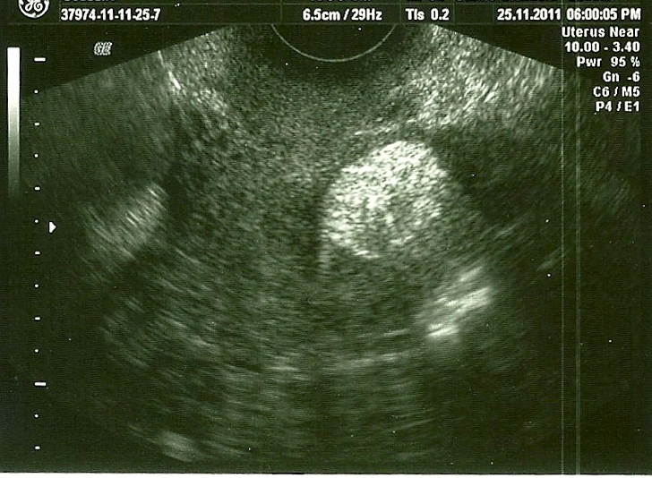
Figure 1:
TSV ultrasound showing a typical hyperechogenic intramural nodule of 2 cm.