


1. Introduction
The athlete’s heart syndrome refers to the electro-morphological remodeling that occurs to varying degrees depending upon the sporting discipline [1]. Regular participation in intensive physical exercise is associated with central as well as peripheral cardiovascular adaptations that facilitate the generation of a large and sustained cardiac output and enhance the extraction of oxygen from exercising muscle for aerobic glycolysis [2]. Sports are classified according to their types, dynamic (isotonic) or static (isometric). Dynamic exercise involves changes in muscle length and joint movement with rhythmic contractions which develop a relatively small intramuscular force. Static exercise induces development of a large intramuscular force with little or no change in muscle length or joint movement. These two types of exercises should be thought of as the two opposite poles of a continuum, with most physical activities involving both static and dynamic components [3].
For years, it has been believed that predominantly isotonic (aerobic) training leads to more significant changes in LV cavity dilatation, wall thickness, and mass. This is in contrast to athletic activities in which the training is predominantly isometric (strength) in nature, like weight lifting and wrestling, in which there may be only increased wall thickness [2]. However, a recent study showed that the most extreme increases in LV wall thickness had been observed in those athletes training in rowing and cycling. Of note, strength training was associated with only a mild increase in wall thicknesses (although often disproportionate to cavity size), whereas absolute values uncorrected for body surface area usually remained well within the accepted normal range (≤12 mm) [4].
Intense endurance athletic activity is prevalent globally, ranging from recreational, extreme sports to elite athletic activity. The term elite athlete refers to an individual who is currently or has previously competed as a varsity player (individual or team), a professional player or a national or international level player. Furthermore, extreme sports (also called action sports and adventurous sports) is a popular term for certain activities perceived as having a high level of inherent danger but do not necessarily require an intense training program. These activities often involve speed, height, a high level of physical exertion, and highly specialized gear. Finally, recreational sports are sports whose main purpose is maintaining and improving cardiovascular fitness.
While the definition of elite activity is broad, its intention is to reflect competitive sports. There has been an intensive research effort in the cardiovascular effects of amount and type of athletic activity. Echocardiography has been used extensively to assess the athlete’s heart for this purpose [5-7].
There are numerous important structural and functional features that characterize the athlete`s heart. In this review we will discuss some of structural and functional changes, especially of the right ventricle of individuals engaging in extreme athletic activity. We will also mention novel parameters such as speckle tracking and strain/ strain rate imaging. In addition, we will describe the acute and chronic features of cardiac echocardiography and cardiac MRI related to athletic activity as well as the role of imaging in athlete’s heart evaluation. The purpose of the paper is to present echo parameters in elite sports population that are common in order to facilitate better interpretation of their echo readings that may not be necessarily a distinct digression from these common features such as increase end-diastolic diameter of the left ventricle, left ventricular thickness, relative left ventricular thickness compared to end-diastolic inner diameter, and dilated left atrium.
The normal cardiac volume is in the range of 10-12 mL/kg in men and 9-11 mL/kg in women. Athlete’s heart is defined as a heart volume more than 12 mL/kg body weight in women and more than 13 mL/ kg body weight in men. It can be up to a maximum of 19 and 20 mL/ kg, respectively [7-13].
Typically, healthy athletes have a normal resting systolic function while those with athlete’s heart may have a low-normal range or even slightly below it [14,15]. The stroke volume is typically normal in this group [8,9,14,15]. Stress echocardiography can reveal a normal augmentation of LV contractility [14]. In addition, diastolic function is normal or in the high-normal range in this group [10,14].
2. Methodology
A systematic search for echocardiography and athletic activity was performed using PubMed. The study group was focused to age 18 years and older individuals, both females and males, and the sports activity, often referred to as elite, was focused to competitive sports level on both national and international levels. Activities include endurance and strength-trained athletes. For this review, a comprehensive search of relevant databases from 1983 to 2016 was performed. Keywords were chosen according to pertinence and probability of being found; including athletes heart, athletes, heart, echocardiography, right ventricle, right ventricular morphology, endurance athletes, arrhythmogenic right ventricular dysplasia, cardiac remodeling, exercise echocardiography, speckle tracking, Tissue Doppler, Longitudinal Peak Systolic Strain, Strain Myocardial Imaging, and sudden cardiac death were utilized to search for relevant articles. The purpose of this comprehensive review is to present echo parameters in elite sports that can facilitate better interpretation of their echo readings that do not necessarily reflect a digression from their expected normal values and therefore would not a carry a clinical burden. Echo in athletes is a relatively narrow research field, defined by the amount and breadth of the studies; therefore most of the relevant papers to the best of our knowledge were included herein.
We excluded papers that focused primarily on other imaging modalities rather than echo within the same paper. In this systematic review, the PRISMA checklist was narrowed due to the paucity of major studies of echocardiography in athletes. Therefore, the exclusion and inclusion criteria as noted above were limited to the above parameters.
3. Structural and Functional Remodeling
Endurance training has both short-term and long-term effects on the cardiovascular system. The short-term effects include increase in cardiac output, stroke volume, oxygen consumption and systolic blood pressure along with a decrease in peripheral vascular resistance. The long-term effects of endurance training on the cardiovascular system causes an increase of oxygen uptake, thus increasing the volume load on the LV and RV. The increase in LV volume, specifically an increase in end diastolic causes an increase in LV thickness. Dilation of the left atrium is also a subsequent response to the increase in LV volume.
Moro et al., using Doppler echocardiography, assessed cardiac structural, systolic, and diastolic function parameters in 53 athletes and 36 non-athlete controls.
Several interesting results were observed. First, athletes, compared to those not engaged in athletic activity, showed higher left atrial volume, left ventricular (LV) thickness, and LV and right ventricular (RV) diastolic diameters. Cyclists showed a higher left atrium and LV diastolic diameter when compared to runners. The RV diastolic diameter was higher in cyclists when compared to soccer players [16].
LA and RA remodeling was studied in athletes to determine if the changes are physiological and what is the limitation of that. RA and LA enlargement with normal filling pressures and low bi-atrial stiffness are collectively considered as a feature of the heart athletes. Left atrial volume index and right atrial volume index significantly increased after training in female athletes. Moreover, LA and RA global peak, atrial longitudinal strain and peak atrial contraction strain decreased; however less significant in RA than LA, with no significant changes in biventricular E/e' ratio. These changes are considered as a physiological acclimatization to exercise [17,18].
LV mass index was higher in athletes compared to those not engaged in athletic activity; with cyclists having higher values compared runners and soccer players in this area. Interestingly, the LV systolic function did not differ significantly between the different groups. Several characteristics of diastolic function were evaluated as well, the E/A ratio was higher in cyclists compared to controls. No difference in LV E/E' ratio was observed. The RV systolic function was evaluated using tissue Doppler imaging. Here, the RV systolic function was higher in cyclists and soccer players than runners. No conclusive differences in RV diastolic function were observed in the study. The authors concluded that soccer players, runners and cyclists had remodeling of left and right ventricular structures when compared to controls. The remodeling was more intense in cyclists than runners and soccer players [16].
There are several longitudinal studies that investigate the midterm change in heart champers in response to training. They found momentous increase in LV mass, LVEDV and LVESV with meager increase in LV Global Longitudinal Strain after training, which indicates physiological acclimatization of LV in response to training [19]. Furthermore, Baddish et al. investigates longitudinal changes in ventricular parameters with endurance training and concludes that the developed biventricular dilatation with enhanced diastolic function is connected to training related adaptation [20]. On another study that interrogates mid-term atrial changes in response to training in preadolescent age shows increase in RA and LA sizes with preserved function that, indeed, imply physiological adaptation to exercise conditioning [21].
4. Right Ventricular Morphology and Function
In a study by D’Andrea et al., a total of 430 top-level athletes (220 endurance-trained athletes and 210 strength-trained athletes) underwent transthoracic echocardiography exams using real-time three-dimensional (3D) measurements which included serial shortaxis reconstructions of the right ventricle (RV) and assessments of RV volume. Calculations of the end-diastolic and end-systolic RV volumes and ejection fraction has been performed using Simpson`s method [22].
Several important results relevant to endurance-trained athletes were demonstrated. First, all RV diameters and 3D volumes and all transmitral and transtricuspid Doppler indexes were greater in these athletes. Second, RV end-diastolic volume was significantly greater in endurance trained athletes compared to strength-trained athletes and controls. Third, left ventricular stroke volume, cardiac index, and pulmonary artery systolic pressure were greater in the endurance trained group. Furthermore, using univariate analysis, D’Andrea et al. showed that 3D RV end-diastolic volume was significantly associated with advanced age, male gender, duration of training, endurance training, increased left ventricular stroke volume, and pulmonary artery systolic pressure. Moreover, D’Andrea et al. showed that in the overall study population, the type and duration of training (P < .01), pulmonary artery systolic pressure (P < .01), and left ventricular stroke volume (P < .001) were the only independent predictors of RV end-diastolic volume.
La Gerche et al. demonstrated that intense endurance exercise causes acute dysfunction of the RV; it has been proposed that limited pre-event training may constitute a relative risk for cardiac injury. However, RV arrhythmias and chronic RV remodeling have most commonly been described among highly trained athletes who frequently compete in events of longer duration than a marathon, in contrast, acute changes in LV function were not observed. Although short-term recovery does appear to be complete, chronic structural changes and reduced RV function has been evident in some of the most, well-trained athletes. Clinical significance of long term effects has yet to be elucidated [56].
There has been evidence that the high wall stress of the RV during intense sports increased wall stress may lead to cellular disruption and leaking of cardiac enzymes and may even result in transient RV dilatation and dysfunction. B-type natriuretic peptide (BNP) elevations were observed after a race, which may reflect elevated LV and RV filling pressures [56]. It has been suggested that repetitive elevations in wall stress along with myocyte damage may lead to chronic remodeling. This may induce a pro-arrhythmic state [6].
La Gerche et al. have demonstrated a lower than expected rates of desmosome gene mutations in athletes. The authors suggest that arrhythmogenic right ventricular cardiomyopathy (ARVC) phenotype may be acquired through intense exercise. No identifiable genetic predisposition for this particular phenotype was shown by the investigators [23-25]. However, while five causative desmosome genes have been identified in ARVC, only 30% to 50% of patients with ARVC have one of these abnormal genes. Marcus et al. suggested that other, yet to be identified, genes may also be involved [23,25].
Vitarelli et al. in a novel study assessed the biventricular function in athlete’s hearts using three-dimensional echocardiography and strain imaging [26]. They analyzed endurance trained athletes, strength trained athletes, and martial artists (who are a mix of endurance and strength trained athletes). They found that endurance athletes develop eccentric hypertrophy due to an increase in LV thickness and volume overload, and the strength trained athletes developed concentric hypertrophy with relative LV thickness due to high systolic systemic blood pressure. The martial artist, or the mixed trained athletes had some dimensions of eccentric and concentric hypertrophy, and all athletes demonstrated a decrease in RV longitudinal strain. It has been suggested that different regions within the RV are more susceptible to RV dilation causes ventricular arrhythmias, thus certain regions in the RV adapt differently with extreme exercise [27]. A meta-analysis showed RV end-diastolic volume, mass and LA diameter were higher in endurance athletes than controls [28]. One study compared RV function and size between male and female endurance athletes. Both males and females displayed functional and structural cardiac remodeling; however, the male athletes had a larger RV size and less RV longitudinal strain [29]. Although the RV volume and strain is increased in athletes, at rest the RV ejection fraction usually subsides. In comparison to patients diagnosed with ARVD the RV strain does not change during rest [30].
Unfortunately, the current values about RV parameters in athletic heart is variable and overestimating RV dimensions in athletes. Furthermore, the revised recommendations of American Society of Echocardiography do not include cardiac adaptation in athletes. D’Ascenzi et al. meta-analysis for Normative Reference Values of Right Heart in Competitive Athletes suggested that male competitive athletes had higher upper limits for RVOT parasternal long-axis, mid-cavity diameter, RA area, and RV basal and RV end-diastolic and end-systolic areas. In contrary, the values in athletes were almost the same to normal subjects for RVOT parasternal short-axis. They also suggested different values for females and different ethnicities, however, due to limited studies involving these variables and limited number of participants causes ambiguity in the results of these groups [31]. However, from the available literature, the effect of ethnicity on distinguishing cardiac adaptation in athletic heart from cardiac diseases especially ARVC is minimal [32]. In another study [33], Olympic male athlete playing the major impact found to have values above the upper limit of revised Task Force reference values in about 30%, that is why they recommend 95th percentile as a cutoff value and, moreover, using only major criteria task force for diagnosing ARVC [34].
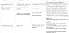
5. Common (and Normal) vs. Uncommon (and Abnormal) Echocardiographic Findings in Athletes
Echocardiography is inexpensive, non-invasive diagnostic tool to differentiate between cardiac adaptation in athletes and cardiac pathology. The values are to differentiate athletic heart from early stages of common diseases that can cause sudden cardiac death. Nonetheless, there is no single optimal test to completely exclude heart disease in athletes, but it should be based on clinical examination, family history, Electrocardiogram and Echocardiography or/ and other imaging modalities. In comparison with hypertrophic cardiomyopathy, LVEDD is >54 mm in athletic heart vs. <45 in hypertrophic cardiomyopathy; LV volume/LV mass, E´mitral lateral annulus, LV radial and circumferential Strain are normal in athletic heart and reduced in hypertrophic cardiomyopathy. In LV noncompaction cardiomyopathy, trabeculation location is apical with reduced or normal values in E´mitral lateral annulus, LV Global Longitudinal Strain at rest and during effort. On the other hand, athletic heart has trabeculations at mid-cavity with normal or supernormal E´mitral lateral annulus, LV Global Longitudinal Strain at rest and during effort. Lastly, ARVC has early RVOT dilatation with motion abnormality and >1 Ratio RV/LV volumes vs. global dilatation without motion abnormality and <1 ratio [35].
6. Exercise Echocardiography
Echocardiography performed before and immediately after exhausting endurance exercise, or endurance competitions showed a mild impairment of diastolic function, and sometimes of systolic function [6,36-38]. The echocardiogram demonstrates acute exerciseinduced functional changes in healthy athletes have shown to be transient, mild and questionable clinical significance [8,37-39].
Stress echocardiography is also useful to augment the endurance athlete’s systolic function.
7. Regional Myocardial Function by Speckle Tracking and Tissue Doppler
Longitudinal Peak Systolic Strain (LPSS) is important for assessment of regional myocardial contractility. This is relevant in athletes in whom regular training can assist in evaluating changes in both ventricles and atria at different work-loads [7,40].
Tissue Doppler and with two- and three-dimensional speckle tracking in athletes show a regional systolic and diastolic left and right ventricular myocardial function in the low-to high-normal range as is left and right atrial function [10,13,41-50]. In addition, Tissue Doppler Imaging (TDI) and Strain Myocardial Imaging (SI) have the have been used to evaluate systolic and diastolic myocardial function in athletes. Speckle tracking techniques have the capability of assessing the LPSS of LV and RV from B-mode imaging in real time. A uniform accuracy in all segments can be achieved; however, dissimilar segmental contributions of the two chambers to overall myocardial contraction may be observed with this approach [7,40].
In a study conducted by Stefani et al, 32 subjects (20 athletes and 18 controls) were evaluated with echocardiography at rest and after a Hand Grip (HG) stress. A four-chamber-view image for LPSS parameter measurements with Speckle Tracking analysis in the basal and medium-apical segments of the two ventricles, at rest and after HG was performed. It was shown that only in athletes, after isometric stress, and increasing afterload, the two ventricles show particular myocardial deformation properties of the regions around the apex wherein the curvature of the wall is particularly more marked [51].
Transthoracic echocardiography, M-mode, 2-D measurements, Doppler derived mitral-tricuspid annular velocities, reconstructed spectral pulsed wave tissue Doppler velocities, strain and strain rate imaging have been used by Tümüklü et al. to evaluate seven myocardial segments in 24 professional soccer and 20 adjusted controls. The study had comparable body surface area, blood pressure and heart rate between the two studied groups. The soccer players group had significantly increased LV mass, mass index, endsystolic and end-diastolic volume, left atrial diameter and decreased transmitral diastolic late velocity. The mass index was due to a higher wall thickness and end-diastolic diameter. The TDI analysis of athletes showed significantly increased mitral annulus septal TDI peak early diastolic(e) velocity, lateral TDI peak e velocity and lateral TDI e/a ratio compared with controls. In addition, the SI analysis of athletes showed increased values of mid septal walls and mid lateral walls peak systolic strain rate values [40].
Tümüklü et al. showed that TDI can be used in athletes such as professional football players to detect, morphologic alteration in left ventricle and left atrium and improvement in left ventricle diastolic function. Furthermore, strain rate imaging can detect subtle changes in systolic left ventricular function in these athletes that cannot be determined using standard echocardiographic parameters and methodologies [40].
Indeed, the 3-dimensional contraction as well as the relaxation of the right and left ventricles is complex. It includes deformation in longitudinal, circumferential and radial planes, as well as deformations in oblique planes (visualized as shear deformations) and twisting [52]. These 3-D deformations can be more accurately studied by MRI that is not limited by imaging planes. Due to the complex pattern of deformation, the basic parameters that describe the myocardial function (i.e right ventricular ejection fraction, TAPSE, RV performance index [Tei index], and RV fractional area change) are not sufficient [51].
Strain by speckle tracking is preferred method since it is not angle dependent and shows a good reproducibility, despite lower temporal resolution. Right ventricular strain and strain rate, both radial and longitudinal, were measured in children using tissue Doppler imaging with a good reproducibility. Longitudinal strains were maximal in the mid part of the RV free wall (peak systolic strain rate was: -2.8 ± 0.7 s-1, peak systolic strain was -45% ± 13%) [53] (This shows normal values in young individuals).
Overall, the free wall right ventricular longitudinal strain was suggested as (-) 27 ± 2%, by a meta-analysis done by Fine et al. [54]. RV free wall longitudinal strain of > (-) 20% was considered abnormal. Right ventricular strain has been found to be a powerful prognostic parameter in patients with pulmonary hypertension, pulmonary embolism, heart failure (with either reduced or with preserved ejection fraction), coronary artery disease. It correlates better with invasive hemodynamic parameters and with cardiac MRI than other parameters (i.e RV fractional area change, TAPSE or peak systolic velocity (S` by tissue Doppler). Importantly and relevant to young athletes, it may show subtle abnormalities in patient with early stages of arrhythmogenic RV cardiomyopathy. 3-D imaging of the right ventricle is an emerging area. It is particularly useful in imaging of the right ventricle due to its complex geometry [55].
8. Comparative Imaging by Echocardiography and MRI
Spence et al. compared echocardiography and cardiac MRI in twenty-two young men randomly assigned to undertake a supervised and intensive endurance or resistance exercise-training program for 24 weeks. It was demonstrated that echocardiography and cardiac MRI showed findings that differed with respect to changes in cardiac size and volume following exercise training. The following were evaluated before and after training: left ventricular (LV) mass, LV end-diastolic volume, internal cavity dimensions, and wall thicknesses [56].
Several important results were demonstrated by Spence et al. First, baseline pooled data for all cardiac parameters were significantly different between imaging methods; however, LV mass (r = 0.756, P < 0.001) and volumes (LV end-diastolic volume, r = 0.792, P < 0.001) were highly correlated between cardiac MRI and echocardiography. Second, changes in cardiac morphology during exercise training measured by echo yielded different and sometime contradictory results compared to MRI. This was particularly evident with posterior wall thickness measurements which increased by 8.3% (P < 0.05) when assessed using echocardiography. It decreased by 2% when using cardiac MRI [56].
In another study, new single beat 3D echocardiography (sb3DE) was compared to cardiac magnetic resonance imaging MRI for measuring right ventricular (RV) dimension and function immediately after a 30 km run. In this study of 21 non-elite male marathon runners, sb3DE was performed and compared with Cardiac MRI.
RV dimensions measured by cardiac MRI were consistently larger when compared to measurements by sb3DE. However, sb3DE and Cardiac MRI did not show significant difference in the RV ejection fraction before and after exercise. Moreover, cardiac MRI showed a significant decrease in RV dimensions while sb3DE measurements did show significant a decrease of RV volumes [57].
Magnetic resonance tissue tagging allows tracking regional myocardial deformation throughout the cardiac cycle, a potentially very useful tool for athletic activity. Using MR tagging, Stuber et al. assessed changes in cardiac torsion and diastolic relaxation of the left ventricle in patients with, healthy control subjects and championship rowers having physiological volume-overload hypertrophy [58]. In the healthy control subjects, a systolic wringing motion was seen with the left ventricle. This motion had a characteristic counterclockwise rotation at the apex and a clockwise rotation at the base; in addition, the apical untwisting motion preceding diastolic filling was observed. In the athletes’ group, LV torsion and untwisting did not change when compared to the control groups [58].
Other studies showed that the myocardial untwisting and untwisting rate were augmented in athletes, thus facilitating early diastolic filling. This has been suggested to occur through the mechanism of suction [59,60].
LV torsion and LW untwisting is enhanced with exercise and appears to have to be associated with pressure gradients in the base to apex. This phenomenon assists in LV filling, whereas this is absent in hypertrophic cardiomyopathy [61].
Indeed, imaging of the right ventricle is challenging, due to the geometric structure of the right ventricle. Moreover, its function including contraction and relaxation is difficult to image and evaluate by echo. Therefore, the gold standard is indeed- cardiac MRI. The reason is that MRI provides a superb 3-D imaging of the RV not restricted by acoustic windows and it has an excellent image quality. Limitations are cost, availability and expertise in interpreting these images. Also, claustrophobia is a limitation. Finally, the temporal resolution of echo is better than cardiac MRI. There is a significant progress analyzing the right ventricle by echo, especially with the development of 3-D echo imaging. Still imaging quality can be challenging at times.
9. Conclusion
In summary, echocardiography can be a useful tool to evaluate athletes in various sports. Table 1 depicts and summarizes the studies done and shows the measured components and findings across the different echocardiography modes and techniques among different athletes. When compared to MRI, echocardiography showed similarities in cardiac morphology data evaluations [56]. The reason that we decided to emphasize the right ventricle is that, in contrast to the left ventricle, the research and the number of publications concerning the right ventricle are relatively limited. Yet, the right ventricle is a very important in the pathophysiology of exercise and may be profoundly affected by heavy exertion (including extreme endurance and resistance exercise) as was mentioned in our review. There are important diseases that preferentially affect the right ventricle (of those, the most well know is arrhythmogenic right ventricular dysplasia, as well as other diseases). Imaging of the right ventricle is challenging due to its complex geometric structure and the pattern of its contraction. MRI and 3-D echo imaging as well as strain imaging are new developments that may help understanding these complex features of the right ventricle. We wanted to bring to the attention of the readers the challenges the progress in imaging of the right ventricle. There are yet additional important questions to be answered as the exact role of echo in monitoring athletes, and its prognostic value. In addition, the effects of performance enhancing drugs and reverse remodeling after discontinuing extreme athletic activity have not been thoroughly studied. The answers to these questions can only be answered with research collaboration between the cardiac community and national/ international sport societies.

Competing Interests
The authors declare that they have no competing interests.
Acknowledgments
This work is supported, in part, by the efforts of Dr. Moro O. Salifu M.D., M.P.H., M.B.A., M.A.C.P., Professor and Chairman of Medicine through NIH Grant number S21MD012474.