


1. Introduction
Chronic obstructive pulmonary disease (COPD) is an important cause of morbidity and mortality around the world. Estimates show that in 2030 COPD will become the third leading cause of death worldwide [1]. Comorbidities in COPD have been studied extensively as COPD patients frequently suffer from comorbidities such as cardiovascular and cerebrovascular disease, lung cancer and diabetes. Research studying the impact of multimorbidities facing COPD patients is necessary as it drives the development of better intervention and diagnostic strategies. Comorbidities have a significant impact on mortality, termed by Divo et al. as the “comorbidome” [1]. There is evidence that comorbidities increase the risk for exacerbations, reduce health status, and increase the risk of mortality [3,4].
The diagnosis of heart failure with preserved ejection fraction (HFpEF) is more challenging than the diagnosis of heart failure with reduced ejection fraction (HFrEF) [5]. According to European Society of Cardiology, patients with HFpEF have an increase in LV wall thickness that results increased filling pressures. The precursor to HFpEF is abnormal left ventricular relaxation, termed left ventricular diastolic dysfunction (LVDD) [5,6]. One half of heart failure cases are attributed to HFpEF [7].
LVDD involves functional abnormalities including decreased myocyte relaxation, and decreased filling or distensibility of the LV, regardless of whether the left ventricular ejection fraction (LVEF) is normal or abnormal or whether the patient is symptomatic or not. LVDD has been associated with coronary artery disease (CAD), Diabetes (DM) and obesity [8], hypertension, and rheumatoid Arthritis (RA) [9]. Consideration for LVDD is an important part in the evaluation of a patient with dyspnea. The echocardiogram is the key diagnostic modality for identifying diagnostic dysfunction. It can also identify concurrent disorders and classify diastolic dysfunction by grades. The importance of identifying LVDD in the COPD population is because it can contribute to dyspnea and lead to heart failure. Both COPD and heart failure exacerbations can present similarly posing a challenge in distinguishing one from the other, even more challenging is that symptoms of one may be incremental to the other.
2. Methods
2.1 Literature search and data sources
An electronic search was performed by two (2) investigators using the PUBMED, Medline, and Cochrane Library databases at the library of the University Hospital of Brooklyn. We used a time (from 2000 to 2018) and language restricted (English) search strategy to identify 4912 articles. Five sets of search terms were used to ensure an adequate and comprehensive literature review. These included “Doppler and COPD,” “Diastolic and COPD,” “Diastolic dysfunction and COPD,” “Atrial and COPD,” and “Ventricular and COPD.” After title and abstract review, the bibliographies of studies that met our inclusion criteria were examined for suitable additional literature. We excluded studies that were not published to maintain a high-quality meta-analysis.
2.2 Study selection and quality assessment
The inclusion was limited to studies that were (1) case-control in design, (2) compared transthoracic echocardiographic (TTE) parameters in COPD patients to non-COPD patients and (3) performed after January 2000. For case control studies that did not include the relevant TTE parameters, the corresponding authors were contacted to provide such data. When no response was attained, the studies were left out of the data pool. No study was excluded based on the size of the study population, but duplicate articles were excluded. For each study, we applied the Newcastle-Ottawa Scale to assess its quality. The quality scale included three components: selection of the study groups with points ranging from 0 to 4; compatibility of the study groups, with points from 0 to 2; and ascertainment of study groups exposure, points from 0 to 3. The maximum for each study is 9, studies with less than 5 points carry the high risk of bias and were excluded (N=2).
2.3 TTE parameters
In 2016, the ASE/EACVI published an updated guideline for the assessment of LVDD using echocardiography. Their recommendation supports a comprehensive study observing both transmitral inflow parameters and tissue doppler parameters. Various parameters they recommend obtaining include annular e prime velocity: septal e’< 7 cm/sec and/or lateral e’< 10 cm/sec, average E/e’ ratio > 14, LA volume index > 34 mL/m2, and peak TR velocity > 2.8 m/sec. Many other parameters have diagnostic and prognostic utility [17]. All but one study in our analysis was published prior to these recommendations. For the purpose of this analysis, studies reporting any LVDD parameters were included in this meta-analysis. The TTE parameters that were most commonly used in the studies included isovolumetric relaxation time (IVRT, msec), E/A ratio, transmitral A wave velocity (meters/second), lateral E’ velocity (meters/second), E-Wave peak velocity (Epv) (meters/second), and deceleration time (DT, msec). Many of these LVDD parameters change in different directions with increasing diastolic dysfunction grade, highlighting the difficulty in diagnosing LVDD.
2.4 Data extraction
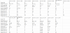
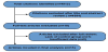
A total of 4,892 studies were excluded for not meeting the inclusion criteria. Eighteen (18) studies meet the inclusion criteria and were reviewed in full; 1 article was removed due to unclear findings, and an additional 2 were removed because the results for their control groups were unclear. Two (2) investigators extracted the data from the fifteen (15) included studies [18-32] with the pooled data represented in tables 1 and 2. Patient characteristics including age, BMI and comorbidity status for each cohort of the individual studies were extracted (Table 3), as well as TTE parameters. The Newcastle-Ottawa Scale for case-control studies was used to assess the quality of the selected articles.
2.5 Statistical analysis
The meta-analysis was performed using Review Manager, version 5.3 (Cochrane Collaboration). Mean differences were evaluated along with 95% confidence intervals (95% CIs). Each included echocardiographic parameter was summarized using the randomeffects model. Heterogeneity, Cochran’s Q, tau-squared test, and I2 index were assessed for each study.
3. Results
There were 1,403 subjects, of which 706 had COPD. In all studies, the diagnosis of COPD was made according to the guidelines of the Global Initiative for Chronic Obstructive Lung Disease (GOLD) criteria.
All of the studies were age and sex-matched. Many of the following were excluded from the studies: 1. Subjects with asthma, hypoxia, pneumonia or patients in acute exacerbation period; 2. Subjects with DM, CAD, hypertension, valvular pathology, left ventricular systolic dysfunction (LVSD) or arrhythmia; 3. Subjects with diffuse lung parenchymal disease, lung malignancies; 4. Subjects with renal failure. Three studies included patients with confounding comorbidities [22,25,26]. The key points of the individual studies are represented in the Table 1. The demographic information for each study can be found in Table 3.
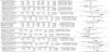
Our meta-analysis found the prevalence of LVDD echocardiographic parameters were higher among COPD subjects versus the control group. We analyzed the most commonly represented LVDD parameters. Patients with COPD had prolonged IVRT (mean difference 20.84 [95% CI 12.21, 29.47]; P< 0.00001), lower E/A ratio (mean difference - 0.24 [95% CI -0.34, 00.14]; P < 0.00001), higher transmitral A wave peak velocity (Apv) (mean difference 11.71 [95% CI 4.80, 18.62]; P< 0.00001), higher E/e’ ratio (mean difference 1.88 [95% CI 1.23, 2.53]; P< 0.00001), lower mitral E wave peak velocity (Epv) (mean difference -8.74 [95% CI -13.63, -3.85]; P< 0.0005), prolonged deceleration time (mean difference 50.24 [95% CI 15.60, 84,89]; P< 0.004), a higher right ventricular end diastolic diameter (RVEDD) (mean difference 8.02 [95% CI 3.45, 12.60]; P< 0.0006) compared to controls. Differences in septal e’ velocity (mean difference -2.69 [95% CI -6.07, 0.69]; P=0.12) and in lateral e’ velocity (mean difference -2.84 [95% CI 5.91, 0.24]; P=0.07) trended towards significance but did not meet our cutoff for statistical significance (p < 0.05).COPD patients had a higher pulmonary arterial pressure (PAP) (mean difference 10.52 [95% CI 3.98, 17.05]; P< 0.002).The details of the individual studies are presented in Table 2.
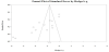
Publication bias was assessed using a funnel-plot. There was no evidence of publication bias in our study. The plot showed a funnel-like shape (Figure 2), with estimated spanning down from the larger trials in both directions with increasing variability. The Figure 2 showed symmetric distributions in both directions and around midline. Given the large heterogeneity, each echocardiographic variable was analyzed separately using the random effects model to calculate the mean difference and 95% CI.
4. Discussion
To our knowledge, this is the first systematic meta-analysis on all case-control studies since the year 2000 looking at diastolic dysfunction in the COPD population. Our review, involving 1,403 patients, reveals that patients with COPD are more likely to have features of LVDD by transmitral inflow patterns and tissue doppler. Compared to the control group, the COPD group had significantly higher E/e’, lower E/A ratio, higher A wave peak velocity, lower E wave peak velocities, and greater IVRT and DT. When taken together, this information suggests that patients with COPD have a greater prevalence of LVDD indices and thus may be prone to develop HFpEF. At least one echocardiographic parameter in all of the 15 studies was statistically different in the COPD group compared to the control group.
Our findings are of clinical significance as concomitant cardiovascular disease (CVD) plays a role in determining the clinical picture in patients with COPD. There is, for example, a 28% increase in cardiovascular mortality for every 10% decrease in FEV1 in COPD [33]. Even when confounders such as exposure to smoke are accounted for, COPD patients have been linked to a 2-3 fold increase in cardiovascular (CV) events [33]. Furthermore, Finkelstein et al., found that the diagnosis of COPD increases the risk of having CVD independently of smoking history, age, gender, lifestyle, and co-morbid risk factors [34]. Interestingly, they also found that COPD reveals the strongest association, greater than that of other cardiovascular diseases, with heart failure [34]. Although the role of coexistent LVDD in CV morbidity and mortality is unknown in the COPD population, in patients with HFpEF rates of hospitalization and death approach those of HFrEF [10,35] .Thus, in the COPD population, HFpEF which may result from LVDD may have an additive detrimental effect [35-36].
Doppler indices to diagnose LVDD are imperfect, and no one parameter has sufficient sensitivity and specificity to diagnose LVDD. However, the more parameters that are abnormal, the more suggestive the diagnosis. The American Society of Echocardiography recommends a comprehensive approach [17]. Evidence of high left ventricular filling pressures are necessary in diagnosing HFpEF [17,37]. A frequently used Doppler parameter is the E/e’ ratio. A higher E/e’ correlates with an elevated left ventricular diastolic pressure, and is the recommended starting point in the diastolic evaluation for patients with normal ejection fraction [17,37]. When the ratio is between 8 and 15, it is important to combine this value with other diastolic parameters. The benefits of the E/e’ ratio is that it is less age dependent, is not influenced by preload and heart rate, is an early marker of LVDD, and provides prognostic information and is very rarely >14 in normal persons [17,37]. Our study found a mean difference in E/e’ of 1.88, greater in the COPD group of patients.
Normal cardiac time intervals indicate normal cardiac function. Thus, time intervals play an important role in assessing for LVDD. As diastolic function declines, early diastolic relaxation proceeds moreslowly, leading to a prolonged IVRT [17,37,38]. The benefit of IVRT is that it provides assessment of the earliest phase of diastole, measuring the interval between aortic valve closure and mitral valve opening [37]. However, it is influenced by tachycardia and arterial pressure and may shorten with increased left a trial pressure, as expected in later stages of LVDD. In our study, there was a mean difference of 20 msec favoring the COPD group, indicating that early diastolic filling is more likely to be impaired in patients with COPD.
Filling patterns such as the E/A ratio and deceleration time (dt) are feasible, reproducible and provide prognostic information [17]. With impaired relaxation, one would expect an increased dt (>220 ms) and an inverse E/A ratio (<1). This pattern has a high specificity for abnormal left ventricular (LV) relaxation and can be seen with normal or increased filling pressures. As diastolic dysfunction worsens in severity, the E/A ratio may normalize, known as pseudonormalization, or even become greater than 2.Similarly, as LVDD worsens LV pressures will reach a point halting early E flow abruptly, causing a decrease in deceleration time. However, impaired relaxation is the earliest step and is expected in all patients with diastolic dysfunction [6]. The E/A ratio in the COPD group in our study was significantly reduced and the deceleration time prolonged, indicating that filling patterns may be consistent with impaired relaxation.
The mechanism(s) of why echo parameters of LVDD are more common in the COPD population are speculative. Possible influences include tachycardia, systemic inflammation, pulmonary hypertension, chronic hypoxemia, chronic hypercapnia, hyperinflation, and right ventricular – left ventricular interaction.
Our study demonstrates that patients with COPD have a tendency towards higher left ventricular filling pressures, as evidenced by the increased E/e’ compared to controls, and worse early diastolic filling as evidenced by lower E/A ratio, lower Epv, higher Apv, and increased IVRT and DT. COPD may lead to pulmonary hypertension, right ventricular hypertrophy and dilation, which may compromise left ventricular filling.
Heart rate (HR) was increased in nearly all of the reviewed COPD groups. Tachycardia particularly effects parameters obtained by doppler echocardiography. Tachycardia can affect E/A ratio, IVRT and DT, however, all but a few studies adjusted their data for HR, or controlled for HR, and yet differences indicating DD persisted. The causes of tachycardia in COPD are manifold; they include side effects of therapy, chronic hypoxia, hypercapnia, and autonomic dysregulation [31,39]. Boussuges et al., found ultrasonographic evidence of decreased LV filling in their COPD group, indicated by a significant decline in the total transmitral VTI [20]. Hyperinflation as a cause of decreased preload in the COPD population has been found in other studies [40-42]. Thus, decreased preload in patients with COPD may also contribute to tachycardia.
Lung hyperinflation in LVDD in the COPD population may be important. Lung hyperinflation and distension may lead to increased stiffness of the parietal pleura, which induces pressure onto the walls of the cardiac fossa. This in turn adds load onto the LV by necessitating conformational changes of the heart during the cardiac cycle [24,31]. This stiffness, imposed upon the heart by the parietal pleura and the cardiac fossa, may be one of the explanations of LVDD in COPD patients irrespective of pulmonary artery pressures.
In addition to lung hyperinflation, the presence of airway obstruction may provoke DD [27]. Hyperinflation and airway obstruction, by increasing positive end-expiratory pressure, may lead to decreased pulmonary vascular compliance, increased right ventricular load, reduced right ventricular stroke volume and, thus, reduced left ventricular filling [21]. Kubota et al. found that E/e’ significantly correlated with patients’ residual volume/total lung capacity ratio [26]. This strengthens the theory that hyperinflation is likely a contributing factor to elevated left ventricular pressures and diastolic dysfunction, and the severity of COPD may relate to severity of LVDD.
Pulmonary hypertension is a common manifestation of long standing or severe COPD, and has been linked to significant effects on morbidity and survival [43,44]. Yilmaz et al. found that LV abnormalities were related to severity of COPD, increased PAP and deterioration of RV function [32]. Suchon et al., also found a strong correlation between the severity of diastolic dysfunction and the severity of the PASP [30]. Sabit et al. found that FEV1 was related to indices of pulmonary hypertension such as pulmonary acceleration time, right ventricular free wall thickness, and myocardial relaxation time. Not surprisingly, pulmonary artery pressure was significantly elevated in the COPD population in our study. Some of same mechanisms which lead to the pulmonary vascular changes in COPD may also contribute to LVDD such as endothelial damage, systemic inflammation and chronic hypoxia causing increased endothelin synthesis and release, and decreased NO and prostacyclin synthesis and release among other mechanisms, and are linked to arterial stiffness [45].
Additionally, chronic hypoxemia may cause calcium transport disturbances leading to myocyte hypoxia and abnormalities of myocardial relaxation [24,46]. The interplay between hypoxemia and interventricular dependence was pointed out in the study by Sabit et al., which found that arterial oxygen tension was related to right ventricular (RV) free wall thickness and myocardial relaxation time [29].
Systemic inflammation is a known contributor to the development of HFpEF. Pro-inflammatory states, such as COPD, lead to an increase in proinflammatory markers such as IL-6 and TNF-α, among others. In the coronary microvasculature this leads to an increase in E-selectin, VCAM and endothelial Reactive oxygen species (ROS) expression and a decrease in nitric oxide (NO) availability [47]. These processes, through further signaling (NO-cGMP-PKG) lead to an increase of collagen deposition via myofibroblasts. Collectively these processes may lead to compromised coronary microvasculature and stiffer myocardial tissue [47]. IL-6 levels were significantly greater in the study performed by Sabit et al. [29]. Additionally, the levels of IL-6 were related to pulmonary acceleration time, LV strain and mitral annular e/a [29].
Arterial stiffness plays an important role in LVDD. In the study by Sabit et al., they found that the COPD group had a 22% greater pulse wave velocity (PWV) [29]. They also found the PWV was related to LVDD parameters such as E/e’; mitral E/A; mitral annular e/a and IVRT. However, they did not find any relationship between PWV and parameters of systolic function. Thus arterial stiffness may manifest in COPD and contribute to LVDD. PWV is a simple and reproducible method of determining arterial stiffness [48]. Arterial stiffness leads to a left ventricular afterload increases because of the earlier return of the reflected arterial wave causing increased left ventricular pressures, increased myocardial oxygen demand, and concomitant reduction in diastolic blood pressures, itself a cause of subendocardial ischemia with resulting myocardial relaxation abnormalities [25,48]. The magnitude of wave reflection is associated with DD [17]. Endothelial dysfunction is a known consequence of COPD seen throughout the disease course, from early to end-stage disease, and is directly linked to irreversible eNOS inhibition by cigarette smoke [44].
Patients with both pulmonary hypertension and LVDD may be more susceptible to dyspnea. The right ventricle is thinner and weaker than the left, meaning it has difficulty adapting to increases in afterload. Generally, the progression of pulmonary hypertension is slow, allowing the right ventricle to adapt to the increases. Infectious exacerbations and exercise can increase the pulmonary pressures significantly, worsening left ventricular filling via interventricular dependence. Patients with LVDD and elevated LV filling pressures cause an increase in pulmonary post-capillary pressures [49]. We found RVEDD was significantly greater in the COPD group, thus the hearts of some patients with COPD undergo conformational changes. This finding helps illustrate the potential link between right ventricular dilation and left ventricular diastolic dysfunction.
The reverse Bernheim phenomenon may explain the influence of the right ventricular volume and or pressure on the left ventricular function [30,50]. The interdependence of the ventricles is due to the left and right chambers being different from one another in sustainable pressures, strength, elasticity and size and yet anatomically bound to one another by the pericardium and their shared interventricular septum. It is postulated that pericardial restraint ensures that with increased pulmonary artery / right ventricular pressures as may occur with COPD, the interventricular septum will deviate into the left ventricle. As a result, there is flattening or reversal of the septal curvature and compression of the left ventricle at end-systole, which may persist in the early diastolic filling period [24,30,49-51]. Our findings indicate that in COPD, LV filling is reduced during early diastole-as indicated by the lower Epv-when septal geometry is compromised, and there is a compensatory increase in late diastolic filling, as indicated by the increased Apv and inverted E/A ratio.
Other factors may contribute to LVDD such as diabetes mellitus, coronary artery disease, hypertension, obesity, and age. The strengthof our study is the analysis of age, sex and co-morbidity matched studies, suggesting COPD as a risk factor for LVDD.
4.1 Clinical implications
Worsening dyspnea in patients with COPD is often thought to be due to progression of the lung disease, however LVDD may lead to increased pulmonary post-capillary pressures and clinically significant HFpEF [30,49]. Our study underscores that COPD patients may have a higher prevalence of LVDD and thus may be more sensitive to preload changes and elevations in pulmonary artery pressure, which may occur from various causes such as arrhythmias and infectious exacerbations [30].
COPD is linked to cardiovascular disease, particularly heart failure [34]. This review underscores the increased risk of LVDD in this group of patients, which may be present even in the early stages of the COPD and often times may go undiagnosed. Fontes-Carvalho showed that COPD patients with LVDD are at increased risk for hospitalization for exacerbation [37]. Reversible ischemic defects may be present at a higher rate in patients with advanced COPD and LVDD thus vigilance for significant coronary disease is prudent [21,52].
Further studies to explore diastolic stress testing in COPD patients may be interesting. A study in non-COPD patents demonstrated that E/e’ > 13 with exercise has incremental prognostic power for ischemic and heart failure events in follow up [53]. Whether similar findings in patients with COPD are seen merits study. Further research highlighting the prevalence of RV dysfunction in COPD patients with LVDD, both on morbidity and mortality, is needed. It is possible that studying left ventricular lusitropy in COPD patients may provide diagnostic and prognostic information, and perhaps help direct therapy.
Competing Interests
The authors declare that they have no competing interests.
Acknowledgments
This work is supported, in part, by the efforts of Dr. Moro O. Salifu M.D., M.P.H., M.B.A., M.A.C.P., Professor and Chairman of Medicine through NIH Grant number S21MD012474.