Profile
International Journal of Clinical Pharmacology & Pharmacotherapy Volume 1 (2016), Article ID 1:IJCPP-121, 6 pages
https://doi.org/10.15344/2456-3501/2016/121
Review Article
DNA Damage Repair and TP53 Gene in Platinum-drug Resistance

Jing Jie Yu

WVU Cancer Institute and Molecular Medicine Core, Department of Biochemistry, School of Medicine and Department of Basic Pharmaceutical Sciences, School of Pharmacy, Robert C. Byrd Health Sciences Center, West Virginia University, Morgantown, WV, USA
Dr. Jing Jie Yu, WVU Cancer Institute, P.O. Box 9300, Morgantown, WV 26506, USA, Tel: (304) 293-8661, Fax: (304) 293-4667; E-mail: jyu@hsc.wvu.edu
26 July 2016; 15 December 2016; 17 December 2016
Yu JJ (2016) DNA Damage Repair and TP53 Gene in Platinumdrug Resistance. Int J Clin Pharmacol Pharmacother 1: 121. doi: https://doi.org/10.15344/2456-3501/2016/121

Abstract

Tumor drug resistance remains a major obstacle in the treatment of cancers. Increased DNA repair is a primary mechanism of acquired resistance to platinum chemotherapy. TP53, Chk2 and ERCC1 of the DNA damage/repair pathway were activated after cells were exposed to cisplatin or dicycloplatin. Overexpression of p53 in wt-p53 (but not p53-deficient) cells doubled Chk2 phosphorylation. p53 knockdown greatly reduced Chk2 phosphorylation, indicating that wild type p53, in response to platinum-drugs, plays a role in the upstream regulation of the DNA-adduct repair pathway and mediates acquired platinum resistance. We strongly suggest that it is important to include p53 mutational status in any p53-involved studies due to the functional differentiation of wt-p53 and p53-mutant.


1. Introduction

Platinum compounds, the DNA-targeting drugs, are effective and have been widely used in clinical settings. However, acquired resistance to platinum-chemotherapy is a major treatment obstacle affecting life-quality of patients and therapeutic outcomes, often leading to treatment failure [1,2]. Studies show that cells treated with genotoxic agents swiftly respond by activating DNA-damage checkpoint response. Two primary pathways are initiated in response to DNA damage. One is mediated by the ATM-Chk2 axis, the other by the ATR-Chk1 axis. The ATM-Chk2 pathway responds primarily to DNA double-strand breaks, whereas the ATR-Chk1 pathway mainly responds to replication-associated DNA lesions [3,4].

Drug-resistance is multifactorial in nature. Its mechanisms involve a complex network of cellular pathways and molecular changes with numerous cross-interactions at different stages. Resistance to platinum-compounds may occur due to alterations of drug influx and efflux; altered activation or metabolism; modified drug-induced damage or drug-targets; and/or evasion of apoptosis [5,6]. To date, however, no successful chemosensitizers or diagnostic/prognostic assays for the prediction of therapy response have been developed.

Transcription factor p53 plays a key role in the DNA damage response to genotoxic stress. Wild type p53 protein has a role in the inhibition of DNA synthesis following DNA damage, suggesting a mechanism for how the loss of wild-type p53 may contribute to tumorigenesis [7,8]. Mutations of p53 have been associated with resistance to platinum-based chemotherapy and shortened survival in ovarian cancer [9].

Checkpoint kinase 2 (Chk2) resides at the heart of the DNA damage/repair pathway and is responsible for the maintenance of mammalian genomic integrity. Studies suggest that Chk2 inhibition in combination with genotoxic agents might have therapeutic value [10-12]. Inhibition of Chk2 expression reduces DNA-damage-induced cell cycle checkpoints and enhances apoptosis in p53-defective HEK- 293 cells [11]. Molecular or genetic targeting of Chk2 prevents the release of survivin from mitochondria and enhances DNA-damage -induced tumor cell apoptosis, thus inhibiting in vivo growth of resistant tumors, providing a rational approach for treatment [13,14].

2. NER Pathway is Associated with Platinum Resistance Phenotype

Nucleotide excision repair (NER) is the critical DNA repair pathway.

It appears to be the major mechanism for removal of platinuminduced DNA adducts, resulting in resistance to platinum drug therapy. NER enzyme-complexes remove bulky, transcription blocking lesions caused by endogenous and environmental insults to DNA, including platinum-induced adducts [15-17]. NER-defective cells are hypersensitive to platinum drugs, and enhanced DNA repair has been implicated in the cisplatin-resistant phenotype [18-20]. The major steps in the NER process are damage recognition, dual-incision of damaged DNA (on both 5' and 3' sides of the lesion), removal of incised nucleotides and deoxyribose, and gap fill-in synthesis [21-26].

3. ERCC1 Expression Predicts Chemosensitivity

ERCC1 is an essential component of the NER pathway, which is the only known mechanism for the removal of intrastrand/interstrand bulky DNA-adducts. Studies indicate that high levels of ERCC1 expression reflect elevated DNA repair capacity and are associated with clinical resistance to platinum-based chemotherapy [18,19,27].

In vitro studies suggest that ERCC1 expression in cisplatinhypersensitive, repair-deficient cells is 50- to 30-fold lower than in platinum-resistant cells [28]. Overexpression of ERCC1 and other NER genes is associated with increased DNA repair activity and clinical resistance to platinum treatment [29,30]. Data from in vitro systems have shown that suppressed ERCC1 expression by siRNA enhances or restores sensitivity of human cancer cells to cisplatin [31]. Other studies also reported that enhanced DNA repair of platinum-DNA adducts or removal of cisplatin-induced interstrand and intrastrand crosslinks has been observed in cisplatin-resistant cell models, and DNA repair inhibitors in some models can potentiate cisplatin cytotoxicity [32-34]. These findings have significant therapeutic implications.

figure 1
Figure 1: Shows the clinical response to cisplatin and the expression of NER genes in tissues of ovarian cancer patients. Panels in A of the two figures show 4 genes: ERCC1, XPA, XPB and CSB expressed at low levels in platinum-sensitive tumors (responders). The same genes expressed at high levels in platinum-resistant patients are shown in the panels in B [27]. These data suggest that clinical resistance to platinum therapy is positively associated with expression of human DNA repair gene ERCC1 and other NER genes.
figure 2
Figure 2: In vitro, the platinum-resistant ovarian cancer A2780/CP70 cells treated with cisplatin showed a 6-fold increase in ERCC1 mRNA level (Figure 2 left panel). This increased mRNA is caused by increased transcription (Figure 2 middle panel) and by prolonged mRNA half-life (Figure 2 right panel). The ERCC1 mRNA half-life is increased or prolonged 8 hours in the cisplatintreated cells compared to untreated control cells [34].

4. Functional Cis-Elements, AP1 and MZF1, in the ERCC1 Promoter Region Responding to Cisplatin Stimulation

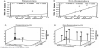
In order to control ERCC1 expression, we performed functional analysis of ERCC1 promoter region by Electrophoresis Mobility Shift Assay and revealed two cis-elements AP1 and MZF1 [35]. In response to cisplatin stimulation, the AP1 site and MZF1 site formed DNAprotein complexes. Furthermore, after cisplatin treatment, binding activities of AP1 increased and binding activities of MZF1 decreased during time course. This suggests that AP1 plays a role as an activator and MZF1 as a repressor. We also measured mRNA expression of AP1 and MZF1 affected by cisplatin and observed that both c-jun and c-fos mRNA levels were increased after cisplatin exposure. In contrast, MZF1 mRNA decreased nearly 75% at 48 hours after cisplatin treatment (data not shown) [34,35].

Put together, our data suggest that within the ERCC1 promoter, the region of -220 to -110 is essential to constitutive ERCC1 expression. And a more forward upstream region containing activator AP1 and repressor MZF1 binding sites is responsible for cisplatin-induced ERCC1 upregulation. In other words, AP1 and MZF1 are the ciselements reacting to cisplatin stimulation. In response to cisplatin treatment, decreased MZF1 and increased AP1 binding activities appear to be the leading mechanism of up-regulation of ERCC1 expression [34,35].

5. ERCC1 SNP Serves as a Prognostic Marker

In 1997, our group discovered a significant single nucleotide polymorphism (SNP) in the ERCC1 gene (GenBank Acc # AF001925). We identified a C to T change at codon 118 in Exon 4 of ERCC1 gene. This change converts a common codon usage to an infrequent codon usage, reducing frequency of use 2-fold. We hypothesized that this SNP would be associated with reduced ERCC1 translation and improved response to platinum chemotherapy [36].

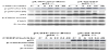
The ERCC1 SNP has been shown by other researchers as an important biomarker associated with platinum sensitivity and predicts better overall survival of patients with several cancers treated with platinum-combination therapy. Specifically, following studies in advanced non-small-cell lung cancer patients, Isla and colleagues concluded that patients homozygous for the ERCC1 118 C allele demonstrated a significantly better survival. They suggest that ERCC1 SNP assessment could be an important component of tailored chemotherapy trials [37]; Ryu et at indicated that median survival time in patients showing C/C genotype of ERCC1 polymorphism was 486 days, which was significantly different from the 281 days of patients with the variant genotype (T/T or C/T; P = 0.0058) [38]. Smith et al. suggested that the C/C genotype at codon 118 may benefit from the combination of platinum and paclitaxel in ovarian cancer patients [39].

6. Wild-Type p53, a Monitor of DNA Damage, Plays a Critical Role in Chemotherapy

In 1994, Lowe and colleagues studied p53 status and the efficacy of chemotherapy in vivo. They found that p53 mutations were detected in resistant or relapse tumor-bearing animals. In response to radiotherapy/chemotherapy, MT-p53 mice responded poorly to the initial treatments, comparing to WT-p53 group, indicating that p53 mutation is associated with treatment resistance and tumor relapse [40]. These findings suggest a basis for the association between p53 mutation and drug resistance/poor prognosis; and between p53 mutation and tumor relapse in patients during chemotherapy.

7. Platinum Drugs Induce p53 Phosphorylation, which Modulates Chk2 Activation

In an investigation of cisplatin-induced molecular signature in cisplatin-sensitive ovarian cancer A2780 cells, we found that several kinases of the DNA-damage repair pathway were activated. One hour after drug exposure we observed phosphorylations of p53 at serine 15 and serine 20, and Chk2 at threonine 68, and increased proteins of ATM, p53, p48 and p21. Of note, cisplatin induced p53 phosphorylation was 12-h earlier than Chk2 phosphorylation, which suggests that Chk2 is activated and regulated by p53 in a wild-type p53 cell model [12]. In another investigation of dicycloplatin-activated DNA-damage response pathway in the same cells, two major factors - p53 and Chk2 - were activated in a manner similar to cisplatininduction, suggesting that the molecular mechanism of dicycloplatin anti-cancer activity is similar to cisplatin [41].

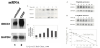
8. Overexpression of p53 Increases Chk2 Phosphorylation in Wild-Type p53 Cells but Not in p53-Null Cells

To investigate our hypothesis that only wild-type p53 phenotype possesses the p53 function, cDNA-transfection was performed in both wild-type p53 A2780 and p53-null SKOV3 cells. Overexpressed p53 gene increased cisplatin-induced Chk2 phosphorylation in the wildtype p53 cell model. It doubled the amount of Chk2 phosphorylation 48-h after drug treatment (Figure 3A). In contrast, western analysis showed no effect on Chk2 phosphorylation by cDNA transfection in p53-null SKOV3 cells (Figure 3B). In other words, transfection of p53 in MT-p53 cells failed to alter Chk2 activation [12].

figure 3
Figure 3: Overexpression of p53 increases chk2 phosphorylation in wild-type p53 cells but not in p53-null cells.

9. Inhibition of p53 by Specific siRNA Inhibits Chk2 Phosphorylation

To confirm the above observations, we performed p53 knock-off siRNA assays and measured Chk2 expression. As shown in Figure 4, in cells not treated with cisplatin, the siRNA to human p53 produced a decrease of phosphorylated Chk2, compared to the nonspecific siRNAtreated control. This decreased level may reflect a constitutive level of activated Chk2 that is normally regulated by p53. Cells transfected by specific siRNA to p53 and treated with cisplatin resulted in a great reduction of phosphorylated Chk2 at Thr-68, suggesting that p53 modulates 68-threonine phosphorylation of Chk2 [12].

figure 4
Figure 4: p53 knock-off siRNA assays and measured Chk2 expression.

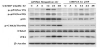
These results indicate that in specific conditions Chk2 activation is regulated by p53 in response to cisplatin treatment in wild-type p53 cells but not in p53-deficient cells. Cells without wild- type p53 could survive, conceivably, via an alternative pathway in response to cisplatin treatment. Therefore, we strongly suggest that any research involving the p53 gene should determine its mutational status due to functional differentiation between wt-p53 and p53-mutant.

figure 5
Figure 5: summarizes the mechanism of cisplatin resistance from our studies: cisplatin activates the DNA damage repair pathway, with marked Chk2 phosphorylation and ERCC1 overexpression. Chk2, the upstream regulator, is modulated by p53. Within the process of cisplatin-induced cell cycle arrest, DNA-adduct repair protein ERCC1 increases and contributes to the increase of cisplatin resistance. Two transcriptional factors AP1 and MZF1 can modulate the ERCC1 expression [42].

Competing Interests

The author declare that there is no competing interests regarding the publication of this article.

Acknowledgments

We acknowledge the Molecular Medicine Core Facility, Mary Babb Randolph Cancer Center, West Virginia University and Sopo-Xingda Pharmaceutical, Inc., Beijing, P.R. China for supporting our studies. Special thanks to Michael D. Mueller for editorial assistance.


References

  1. Kelland L (2007) The resurgence of platinum-based cancer chemotherapy. Nat Rev Cancer 7: 573-584 [CrossRef] [Google Scholar] [PubMed]
  2. Cossa G, Gatti L, Zunino F, Perego P (2009) Strategies to improve the efficacy of platinum compounds. Curr Med Chem 16: 2355-2365 [CrossRef] [Google Scholar] [PubMed]
  3. Bartek J, Lukas J (2003) Chk1 and Chk2 kinases in checkpoint control and cancer. Cancer Cell 3: 421-429 [CrossRef] [Google Scholar] [PubMed]
  4. Pommier Y, Weinstein JN, Aladjem MI, Kohn KW (2006) Chk2 molecular interaction map and rationale for Chk2 inhibitors. Clin Cancer Res 12: 2657-2661 [CrossRef] [Google Scholar] [PubMed]
  5. Longley DB, Johnston PG (2005) Molecular mechanisms of drug resistance. J Pathol 205: 275-292 [CrossRef] [Google Scholar] [PubMed]
  6. Redmond KM, Wilson TR, Johnston PG, Longley DB (2008) Resistance mechanisms to cancer chemotherapy. Front Biosci 13: 5138-5154 [CrossRef] [Google Scholar] [PubMed]
  7. Fojo T (2002) p53 as a therapeutic target: unresolved issues on the road to cancer therapy targeting mutant p53. Drug Resist Updat 5: 209-216 [CrossRef] [Google Scholar] [PubMed]
  8. Kastan MB, Onyekwere O, Sidransky D, Vogelstein B, Craig RW (1991) Participation of p53 protein in the cellular response to DNA damage. Cancer Res 51: 6304-6311 [Google Scholar] [PubMed]
  9. Reles A, Wen WH, Schmider A, Gee C, Runnebaum IB, et al. (2001) Correlation of p53 mutations with resistance to platinum-based chemotherapy and shortened survival in ovarian cancer. Clin Cancer Res 7: 2984-2997 [Google Scholar] [PubMed]
  10. Hirao A, Cheung A, Duncan G, Girard PM, Elia AJ, et al. (2002) Chk2 is a tumor suppressor that regulates apoptosis in both an ataxia telangiectasia mutated (ATM)- dependent and an ATM-independent manner. Mole Cell Biol 22: 6521- 6532 [CrossRef] [Google Scholar] [PubMed]
  11. Yu Q, Rose JH, Zhang H, Pommier Y (2001) Antisense inhibition of Chk2/ hCds1 expression attenuates DNA damage-induced S and G2 checkpoints and enhances apoptotic activity in HEK-293 cells. FEBS Lett 505: 7-12 [CrossRef] [Google Scholar] [PubMed]
  12. Liang X, Guo Y, Figg WD, Fojo AT, Mueller MD, et al. (2011) The Role of Wild-Type p53 in Cisplatin-Induced Chk2 Phosphorylation and the Inhibition of Platinum Resistance with a Chk2 Inhibitor. Chemother Res Pract 2011: 715469 [CrossRef] [Google Scholar] [PubMed]
  13. Antoni L, Sodha N, Collins I, Garrett MD (2007) CHK2 kinase: cancer susceptibility and cancer therapy - two sides of the same coin? Nature Reviews Cancer 7: 925-936 [CrossRef] [Google Scholar] [PubMed]
  14. Ghosh JC, Dohi T, Raskett CM, Kowalik TF, Altieri DC (2006) Activated checkpoint kinase 2 provides a survival signal for tumor cells. Cancer Res 66: 11576-11579 [CrossRef] [Google Scholar] [PubMed]
  15. Huang JC, Svoboda DL, Reardon JT, Sancar A (1992) Human nucleotide excision nuclease removes thymine dimers from DNA by incising the 22nd phosphodiester bond 5' and the 6th phosphodiester bond 3' to the photodimer. Proc Natl Acad Sci U S A 89: 3664-3668 [CrossRef] [Google Scholar] [PubMed]
  16. Moggs JG, Yarema KJ, Essigmann JM, Wood RD (1996) Analysis of incision sites produced by human cell extracts and purified proteins during nucleotide excision repair of a 1,3-intrastrand d(GpTpG)- cisplatin adduct. J Biol Chem 271: 7177-7186 [CrossRef] [Google Scholar] [PubMed]
  17. Furuta T, Ueda T, Aune G, Sarasin A, Kraemer KH, et al. (2002) Transcription-coupled nucleotide excision repair as a determinant of cisplatin sensitivity of human cells. Cancer Res 62: 4899-4902 [Google Scholar] [PubMed]
  18. Reed E (2006) ERCC1 measurements in clinical oncology. N Engl J Med 355: 1054-1055 [CrossRef] [Google Scholar] [PubMed]
  19. Yu JJ, Dabholkar M, Bennett WP, Welsh JA, Mu CJ, et al. (1996) Platinumsensitive and platinum-resistant ovarian cancer tissues show differences in the relationships between mRNA levels of p53, ERCC1 and XPA. Int J Oncol 8: 313-317 [CrossRef] [Google Scholar] [PubMed]
  20. Sood AK, Buller RE (1996) Genomic instability in ovarian cancer: a reassessment using an arbitrarily primed polymerase chain reaction. Oncogene 13: 2499-2504 [Google Scholar] [PubMed]
  21. Sancar A, Tang MS (1993) Nucleotide excision repair. Photochem Photobiol 57: 905-921 [CrossRef] [PubMed]
  22. Matsunaga T, Mu D, Park CH, Reardon JT, Sancar A (1995) Human DNA repair excision nuclease. Analysis of the roles of the subunits involved in dual incisions by using anti-XPG and anti-ERCC1 antibodies. J Biol Chem 270: 20862-20869 [CrossRef] [Google Scholar] [PubMed]
  23. Reardon JT, Sancar A (2002) Molecular anatomy of the human excision nuclease assembled at sites of DNA damage. Mol Cell Biol 22: 5938-5945 [CrossRef] [Google Scholar] [PubMed]
  24. Hoeijmakers JH1 (1993) Nucleotide excision repair I: from E. coli to yeast. Trends Genet 9: 173-177 [CrossRef] [Google Scholar] [PubMed]
  25. Wood RD (1997) Nucleotide excision repair in mammalian cells. J Biol Chem 272: 23465-23468 [CrossRef] [Google Scholar] [PubMed]
  26. Petit C, Sancar A (1999) Nucleotide excision repair: from E. coli to man. Biochimie 81: 15-25 [CrossRef] [Google Scholar] [PubMed]
  27. Dabholkar M, Vionnet J, Bostick-Bruton F, Yu JJ, Reed E (1994) Messenger RNA levels of XPAC and ERCC1 in ovarian cancer tissue correlate with response to platinum-based chemotherapy. J Clin Invest 94: 703-708 [CrossRef] [Google Scholar] [PubMed]
  28. Behrens BC, Hamilton TC, Masuda H, Grotzinger KR, Whang-Peng J, et al. (1987) Characterization of a cis-diamminedichloroplatinum(II)-resistant human ovarian cancer cell line and its use in evaluation of platinum analogues. Cancer Res 47: 414-418 [Google Scholar] [PubMed]
  29. Dabholkar M, Bostick-Bruton F, Weber C, Bohr VA, Egwuagu C, et al. (1992) ERCC1 and ERCC2 expression in malignant tissues from ovarian cancer patients. J Natl Cancer Inst 84: 1512-1517 [CrossRef] [Google Scholar] [PubMed]
  30. Masuda H, Ozols RF, Lai GM, Fojo A, Rothenberg M, et al. (1988) Increased DNA repair as a mechanism of acquired resistance to cisdiamminedichloroplatinum (II) in human ovarian cancer cell lines. Cancer Res 48: 5713-5716 [Google Scholar] [PubMed]
  31. Chang IY, Kim MH, Kim HB, Lee DY, Kim SH, et al. (2005) Small interfering RNA induced suppression of ERCC1 enhances sensitivity of human cancer cells to cisplatin. Biochem Biophysl Resh Commun 327: 225-233 [CrossRef] [Google Scholar] [PubMed]
  32. Parker RJ, Eastman A, Bostick-Bruton F, Reed E (1991) Acquired cisplatin resistance in human ovarian cancer cells is associated with enhanced repair of cisplatin-DNA lesions and reduced drug accumulation. J Clin Invest 87: 772–777 [CrossRef] [Google Scholar] [PubMed]
  33. Johnson SW, Perez RP, Godwin AK, Yeung AT, Handel LM, et al. (1994) Role of platinum-DNA adduct formation and removal in cisplatin resistance in human ovarian cancer cell lines. Biochem Pharmacol 47: 689-697 [CrossRef] [Google Scholar] [PubMed]
  34. Li Q, Gardner K, Zhang L, Tsang B, Bostick-Bruton F, et al. (1998) Cisplatin induction of ERCC1 mRNA expression in A2780/CP70 human ovarian cancer cells. J Biol Chem 273: 23419-23425 [CrossRef] [Google Scholar] [PubMed]
  35. Yan QW, Reed E, Zhong XS, Thornton K, Guo Y, et al. (2006) MZF1 possesses a repressively regulatory function in ERCC1 expression. Biochem Pharmacol 71: 761-771 [CrossRef] [Google Scholar] [PubMed]
  36. Yu JJ, Mu C, Lee KB, Okamoto A, Reed EL, et al. (1997) A nucleotide polymorphism in ERCC1 in human ovarian cancer cell lines and tumor tissues. Mutat Res 382: 13-20 [CrossRef] [Google Scholar] [PubMed]
  37. Isla D, Sarries C, Rosell R, Alonso G, Domine M, et al. (2004) Single nucleotide polymorphisms and outcome in docetaxel–cisplatin-treated advanced non-small-cell lung cancer. Ann Oncol 15: 1194-1203 [CrossRef] [Google Scholar] [PubMed]
  38. Ryu JS, Hong YC, Han HS, Lee JE,Kim S, et al. (2004) Association between polymorphisms of ERCC1 and XPD and survival in non-small-cell lung cancer patients treated with cisplatin combination chemotherapy. Lung Cancer 44: 311-316 [CrossRef] [Google Scholar] [PubMed]
  39. Smith S, Su D, Rigault de la Longrais IA, Schwartz P, Puopolo M, et al. (2007) ERCC1 Genotype and Phenotype in Epithelial Ovarian Cancer Identify Patients Likely to Benefit From Paclitaxel Treatment in Addition to Platinum-Based Therapy. J Clin Oncol 25:5172-5179 [CrossRef] [Google Scholar] [PubMed]
  40. Lowe SW, Bodis S, McClatchey A, Remington L, Ruley HE, et al. (1994) p53 status and the efficacy of cancer therapy in vivo. Science 266: 807- 810 [CrossRef] [Google Scholar] [PubMed]
  41. Yu JJ, Yang XQ, Song QH, Mueller MD, Remick SC (2014) Dicycloplatin, a novel platinum analog in chemotherapy: Synthesis of Chinese preclinical and clinical profile and emerging mechanistic studies. Anticancer Res 34: 455-464 [Google Scholar] [PubMed]
  42. Yu JJ (2009) Unlocking the Molecular Mechanisms of DNA Repair and Platinum Drug Resistance in Cancer Chemotherapy. Current Drug Therapy 4: 19-28 [CrossRef] [Google Scholar]