


1. Introduction
Various factors including the increased numbers of immunocompromised patients (e.g. AIDS and cancer patients), a growing aged population, the increased use of indwelling medical devices and antibiotic misuse have led to the increase in Candida infection incidence. C. glabrata is one of non - C. albicans species which has become of concern because of its increased incidence and ability to develop resistance towards common antifungals [1].
Detailed genetic studiesof C. glabrata are now commonplace thanks to the regulatable promoters which are available to the researchers to induce or repress gene expression in C. glabrata. These promoters, such as tetO, MET3 and PDC1 promoters [2,3] control expression of the gene of interest upon incubation of cells in growth media containing the activating or the repressing substance. The tetregulatable promoteris directed affected by tetracycline,induced by addition of doxycycline to the growth medium with induction being reversed by doxycycline removal. This system has been successfully used forfunctional analysis studies of essential genes with unknown function in C. glabrata [2]. Doxycycline was described to have no detectable effect on global gene expression in Saccharomyces cerevisiae at certain concentration [4]. However, there are no studies that examine the effect of doxycycline on C. glabrata global gene expression.
In mammalian cells doxycycline was shown to be an ERK1/2 inhibitor [5]. Abioinformatic database analysis (Candida Genome Database (CGD)) indicates that Homo sapiens ERK2 shows 48% identity and 65% similarity to the C. glabrata SLT2, the MAPKKKK protein of cell wall integrity mitogen activated protein kinase signaling pathway (CWI MAPK). Based on that, we set out to determine the effect of doxycycline on SLT2 phosphorylation status in C. glabrata and onits survival in general.We believe that research community in the field of yeast molecular genetics may benefit from the conclusions of our studies.
2. Materials and Methods
2.1 Susceptibility of C. glabrata to doxycycline
C. glabrata ATCC90030 and C. glabrata ATCC2001 strainswere used in this study. Susceptibility testing to doxycycline was performed using three methods. The first method was the standardized broth microdilution assay as defined by CLSI (2008) using RPMI1640 medium (Sigma) and C. glabrata ATCC2001. Doxycycline concentrations ranged from 6.2 to 200 μg/ml. After incubation for 24 h at 35ºC the minimum inhibitory concentration was determined visually. Susceptibility was also observed by spotting 5 μl from serial dilutions of both strains onto YEPD agar plates [1% (w/v) yeast extract, 2% (w/v) bacteriological peptone, 2% (w/v) dextrose and 1.5% bacteriological agar (Oxoid)] contain 100 or 200 μg/ml doxycycline.
Thesensitivity of C. glabrata to doxycycline was tested in water as well. High density cells in water (about 3-8 x 10⁶CFU/ml) were treated with different concentrations of doxycycline ranging from 0 to 400 μg/ml. Preparation of C. glabrata cells for doxycycline treatment in water was carried out as described previously in [6]. Doxycycline was dissolved in RPMI1640 medium or added directly to water.
To examine the effect of sorbitol and antioxidants such as ascorbic acid and reduced glutathione on inhibition of 50 μg/mlof doxycycline toxicity in water, 1 M of sorbitol or 10mM of ascorbic acid or reduced glutathione were added to about 3 x 10⁵ CFU/ml of C. glabrata ATCC90030 together with doxycycline.
2.2 Susceptibility of C. glabrata to doxycycline
Detection of SLT2 phosphorylation by using of flow cytometry was carried out using phospho-p44/42 MAPK (Erk1/2) (Thr202/Tyr204) antibody (Cell Signaling Technology). Cells of C. glabrata ATCC2001 from an overnight YEPD plate were used to inoculate YEPD liquid medium and incubated 4-5 h at 30ºC with agitation (200 rpm). Cells were harvested and washed with RPMI1640 liquid medium and resuspended in the same medium at density of about OD600=1.
To examine SLT2 phosphorylation induction by doxycycline, C. glabrata ATCC2001 exponential phase cultures were examined 30 min after addition of 0, 25 or 50 μg/ml doxycycline. Cells were fixed, permeabilised and antigens detected according to the primary antibody manufacturer’s instructions. In brief, one ml samples were collected from each treatment and 100 μl of 37% formaldehyde was added. The samples were incubated at 37ºC for 10 min, chilled on ice for 1 min and centrifuged (9000 rpm) for 1 min. The pellets were resuspended in 100 μl PBS. Permeabilisation was carried out by adding 900 μl of 100% cold methanol (-20ºC) and incubated on ice for 30 min. For immunostaining, cells were harvested and washed with PBS. Pellets were resuspended in blocking buffer by vortexing and incubated for 10 min at room temperature. Then the cells were harvested and resuspended in blocking buffer containing 1:200 diluted phospho-p44/42 MAPK primary antibody(thr202/tyr204; Cell Signaling Technology #9101). Cells were incubated for 1 h at room temperature. After washing with PBS the cells were suspended in 1:1000 diluted anti-rabbit HRP secondary antibody (Sapphire, Australia) in blocking buffer and incubated for 30 min in the dark at room temperature. After three washes with PBS, fluorescence intensity was detected by AmCyan filter in FACS Canto II (BD Biosciences) flow cytometer.
3. Results
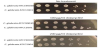
3.1 Susceptibility of C. glabrata to doxycycline in media
The minimum inhibitory concentration of doxycycline to C. glabrata ATCC2001 in RPMI1640 medium was 200 μg/ml. A weak growth was visualized in the microwells which contained 100 μg/ml doxycycline comparing with growth control microwells. Aliquotes of serial dilutions of both strains on YEPD agar containing 0, 100 or 200 μg/ml doxycycline showed no inhibition of growth (Figure 1).
3.2 Susceptibility of C. glabrata to doxycycline in media
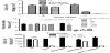
C. glabrata high density cellpopulations (~3 x 10⁶ CFU/ml) of ATCC90030 strain or 8 x 10⁶ CFU/ml of ATCC2001 strain in water began to lose more than 60 and 80%of their viability respectively after treatment with 40 μg/ml or 50 μg/ml of doxycycline (Figure 2a and Figure 2b). The toxicity of doxycycline in water could be partly abrogated (20% loss in viability) by adding 10 mM ascorbic acid but not reduced glutathione (> 80 % loss in viability). Sorbitol also could not save cells from doxycycline toxicity in water (Figure 2c).
3.3 Doxycycline induces SLT2 phosphorylation
Experiments were designed to find whether doxycycline may act as SLT2 inhibitor, and toxicity of doxycycline in water was due to this property. Cells in RPMI1640 were treated with doxycycline and examined for SLT2 phosphorylation. Concentrations of 25, 50 and 100 μg/ml of doxycycline were tested for their ability to inhibit SLT2 phosphorylation. The phosphorylation of SLT2 in C. glabrata ATCC2001 was measured using flow cytometry. Within 30 min of incubation in the lowest concentration of doxycycline (25 μg/ml), phosphorylation of SLT2 protein was detected. The phosphorylation was higher in the cells treated with 50 μg/ml of doxycycline, whereas untreated cells showed no detectable positive events indicating no SLT2 phosphorylation in these cells (Figure 3).

4. Discussion
Doxycycline concentrations of 200 μg/ml inhibited C. glabrata ATCC2001 growth in the MIC assay, while a concentration of one fifth of the MIC of doxycycline was enough to cause loss in viability of this fungus in water. In our previous study>104 CFU/ml of C. glabarat was shown to be able to keep their viability in water over at least three days [6]. This fact has been shown again in this work as 8 x 10⁶ CFU/ ml of C. glabarat ATCC2001and ~ 3 x 10⁵ of C. glabrata ATCC90030 were able to maintain their viability in water.
Many studies depend on spotting serial dilution of yeast on YEPD agar plates to examine the effect of doxycycline on promoter induction [7] or synergy with other compounds [8]. Therefore, the spotting assay was carried out in this study to observe the effect of 100-200 μg/ml of doxycycline on C. glabrata reference strains. Both strains were not sensitive to the highest concentration of doxycycline. However, other yeast species such as C. albicans was susceptible to doxycycline at 200 μg/ml [8] when it was tested by using the spotting assay. A study on S. cerevisiae cells treated with 40 μg/ml of doxycycline showed that there was no phenotypic effect and no significant effect on their global genome transcription levels which allow the use of tetO regulatable promoter system for genetic studies [4]. In our study it is clear that 200 μg/ml of doxycycline in YEPD agar plates did not cause any phenotypic effect on C. glabrata in media but this was not the case when this species was treated with doxycycline in water. It seems that yeast species vary in their response to doxycycline treatment as Fiori et al.,[8] demonstrated by the difference between C. albicans and S. cerevisiae to doxycycline and fluconazole treatment combinations. Growth in the presence of fluconazole and doxycycline was restored by overexpression of ERG11 in S. cerevisiae but not in C. albicans [8].
Since CWIMAPK signaling has been reported to be activated in hypo-osmotic conditions [9] and doxycycline was described as an ERK1/2 inhibitor [5], the homolog of SLT2 in C. glabrata (CGD), the question was asked, could this antibiotic act as a SLT2 inhibitor, leading to cell death in water? The toxicity of doxycycline in water was not abrogated by adding the osmo-stabiliser “sorbitol”. In contrast, staurosporine (a known PKC1 inhibitor) toxicity in water could be abrogated by sorbitol [10]. PKC1 is an upstream kinase of the CWIMAPK signaling pathway. This indicates doxycycline has a target other than CWI-MAPK. Flow cytometry analysis of SLT2 phosphorylation confirmed that doxycycline does not inhibit SLT2 phosphorylation: indeed doxycycline activates SLT2 phosphorylation. The activation of this protein occurred once C. glabrata cells were treated with sub-inhibitory concentrations of doxycycline (50 μg/ml) in RPMI1640 medium.
In summary, the fungicidal effect of doxycycline in water is not due to inhibition of the SLT2 activation. Doxycycline is an iron chelator leading to depletion of intracellular iron [8]. Iron depletion has been proposed to decrease ergosterol content in C. albicans and S. cerevisiae, leading to higher fluidity in cell membranes, with consequent increased passive diffusion [11,12]. Further investigation might include the examination of the effect of iron addition on C. glabrata sensitivity to doxycycline. Water and doxycycline might diffuse passively and cause cell death. The elimination of doxycycline toxicity with ascorbic acid might indicate an interference with the oxidative homeostasis due to iron depletion. In addition, doxycycline was shown to increase the susceptibility of C. albicans to amphotericin when they were treated with 50 to 200 μg/ml of doxycycline [13]. This alteration in susceptibility was suggested to be due to the inhibition of mitochondrial function in C. albicans, which eliminates the diauxic shift [13]. Lack of diauxic shift, or the lack of functional mitochondria alters sterol metabolism, resulting in lower ergosterol levels, consistent with the need for 12 molecules of oxygen to synthesize one molecule of ergosterol [13]. Saccharomyces cerevisiae was shown to require antioxidant function to resist the toxicity of doxycycline [14]: this has been also demonstrated in our study where ascorbic acidprotected against doxycycline toxicity in water. However, it is not clear for us why reduced glutathione, also an antioxidant, did not protect against doxycycline toxicity.
In conclusion this study demonstrates that C. glabratacells are more sensitive to doxycycline in water than in media, and doxycycline isan inducer of SLT2 phosphorylation. Furthermore, this study supports the proposed mechanism of killing of doxycycline in water may be attributed to an alteration of oxidative homeostasis in yeast which alters the cells permeability.
Competing Interests
The authors declare that they have no competing interests.