


1. Introduction
Scedosporium species are emerging human pathogens, responsible for a broad spectrum of infections, including superficial or deep localized diseases in immunocompetent hosts, and disseminated infections in immunocompromised or in near-drowning patients [1]. Scedosporium boydii and Scedosporium apiospermium are mainly associated with infections in temperate climates (e.g., Central Europe), while Scedosporium aurantiacum and Scedosporium prolificans infections are predominant in hot and arid countries (e.g., Spain, Australia) [2]. According to a recent clinical study in Spain, the members of this genus are the second most frequently isolated filamentous fungi from human infections after Aspergillus species [3]. The clinical manifestations of Scedosporium infections are very similar to those of aspergillosis, fusariosis and other hyalohyphomycosis [4]. The proper diagnosis is complicated by the low interspecies diversity and high intraspecies variability, especially within the S. boydii species complex. However, the accurate identification of the causative agent is crucial to find the most effective therapeutic approach, since the antifungal susceptibility profile of Scedosporium species is different from those of hyaline filamentous fungi, and also varies within the genus itself [2]. Another challenge in the treatment is the frequently observed antifungal resistance of the Scedosporium isolates to conventional antifungal agents [1]. In a consequence of these, clinicians have limited options to treat Scedosporium infections. Therefore, new therapeutic strategies are required beside of the currently available ones.
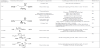
Cysteine and their derivatives have been previously reported to have antifungal activity against different filamentous fungi (Table 1) [5-11]. Previously we proved the antifungal activity of N-acetyl-L-cysteine against Scedosporium species [12]. D-cysteine (DC), L-cysteine (LC), L-cysteine-methyl ester (LCME), N-isobutyryl-D-cysteine (NIDC), and N-isobutyryl-L-cysteine (NILC) showed antifungal effect against species belonging to Mucorales [6]. LC was reported to inhibit spore germination and to cause reduced hyphal growth in different filamentous fungi (e.g. dermatohytes, Aspergillus spp., Fusarium spp.) [7,8,11].
The aims of the present study were (i) to determine the in vitro susceptibility of clinical and environmental Scedosporium isolates to DC, LC and their derivatives, such as LCME, NIDC, and NILC; and (ii) to investigate the in vitro combinations of the most effective cysteine compound with conventional antifungal agents against clinical Scedosporium isolates. developed for the diagnosis of UTIs. Rapid biochemical dipstick tests are available and currently used as predictors of bacterial UTI, but must often be correlated with other testing and clinical information. In many clinical settings, in fact, it has been proved that the dipstick urinalysis leads to many false positive and negative results when compared with the gold standard culture method, demonstrating the low sensitivity and positive predictive value [10]. The Micro Biological Survey method (MBS) is an alternative method for bacterial counting developed and patented by Roma Tre University [11,12]. It is based on a colorimetric survey performed in mono-use disposable reaction vials in which samples can be inoculated without any preliminary treatment. The analysis can be carried out using a thermostatic optical reader that automatically detects the color change. The ease of use of the MBS method has been evaluated in a previous study on food samples demonstrating the possibility to use it anywhere and without the need of an equipped laboratory and specialized personnel [13]. In previous studies carried out on artificially contaminated urine samples, this method has already been proven to be suitable for the evaluation of the bacterial load and the assessment of the susceptibility to a panel of antibiotics [14,15]. The present study has been undertaken to clinically evaluate the performance and operational characteristics of the MBS POCT for the diagnosis and the antibiotic treatment of UTIs. The MBS method was also compared with another biochemical rapid test for UTI diagnosis, namely the urine dipstick assay [16].
2. Materials and Methods
2.1 Strains and culture conditions
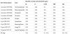
Nine Scedosporium isolates derived from different environmental and clinical sources were involved in this study (Table 2). Prior to the tests, to get the required amount of conidia all the strains were grown on malt extract slants (MEA, Biolab, Hungary) for 2 weeks at 30°C. Susceptibility tests were performed in RPMI 1640 medium (Sigma- Aldrich, USA) supplemented with 0.3 g/l L-glutamine and buffered to pH 7.0 with 0.165 M 4-morpholinopropanesulfonic acid (Sigma- Aldrich, USA) and were incubated at 37°C for 72 hours.
2.2 Microdilution tests
The susceptibility of Scedosporium isolates to cysteine forms and their derivatives were determined following the slightly modified instructions of the Clinical and Laboratory Standards Institute's M38-A2 broth microdilution method [13]. Minimum inhibitory concentration (MIC) was defined as the lowest concentration of a drug which was required for the total growth inhibition of a certain isolate after 72 hours-long incubation. Modifications connected to stock solution and inoculum preparation were detailed previously [14]. We evaluated the antifungal effect of five compounds: DC, LC, LCME, NIDC, and NILC (Sigma-Aldrich, USA). The final drug concentrations in the tests ranged from 64 to 1024 μg/ml.
Drug interactions between LCME and conventional antifungal
agents (i.e., amphotericin B, AMB; caspofungin, CSP; terbinafine,
TRB; and voriconazole, VRC) were investigated against the four
clinical isolates using the checkerboard microdilution method [15].
The final LCME concentrations ranged from 64 to 2048 μg/ml. The
final concentrations of the antifungal drugs were between 0.25 and
128 μg/ml. Fractional inhibitory concentration indexes (FICIs) were
calculated to demonstrate the type of interaction between the paired
combinations of compounds [16]. For these calculations, the MICs
of antifungals have been already determined in a recent study of our
research group [14]. Synergism was defined as FICI≤0.5, indifference
as 0.5
Both single and combination tests were performed in three replicates.
3. Results and Discussion
We evaluated the antifungal effect of two cysteine forms, LC and DC, and their derivatives against nine Scedosporium isolates. The MICs were in the range of 512 - >1024 μg/ml (Table 2). LCME proved to be most effective among the investigated compounds with a MIC range of 512 - >1024 μg/ml. It demonstrated similar antifungal activity against the tested isolates (512 - 1024 μg/ml) except for P. ellipsoidea CBS 301.79 strain where the MIC was >1024 μg/ml. DC, LC and NIDC were ineffective against the Scedosporium isolates in the investigated concentration range (MIC: >1024 μg/ml). While NILC proved to be active against one strain only, S. aurantiacum CBS 116910 (MIC: 1024 μg/ml). Differences between the susceptibilities of environmental and clinical isolates to the investigated compounds were not observed.
The antifungal activity of cysteine and its derivatives against Scedosporium species has not been investigated yet. The MICs observed in this study are comparable to previously reported data against other fungal species. The in vitro inhibitory effect of DC was proved previously against Eutypa lata and different Mucorales species. It caused reduced hyphal growth in a concentration of 0.625 - 10 mM (~ 75 - 1200 μg/ml) [5,6]. The antifungal effect of LC was reported against different filamentous fungi: dermatophytes, Alternaria spp., Aspergillus spp., Eutypa lata, Fusarium spp., and Inonotus obliquus [5-11]. Our previous publication showed the antifungal effect of LCME, NIDC, and NILC against Mucorales species: the MEC values were between 0.625 and 10 mM (~ 100 - 1700 μg/ml at LCME; ~ 60 - 1000 μg/ml at NIDC and NILC) [6]. Beside the reduced hyphal growth, LCME inhibited the germination of sporangiospores, while the presence of NILC in the culture media led to changes in the colony morphology [6]. The mode of action of cysteine and their derivatives has not been clarified yet. Kahlos and Tikka [10] observed that LC reduced the ergosterol production of Inonotus ubliquus. Octave et al. [5] reported the complete structural disorganization of the mycelia of Eutypa lata. According to Daigle and Cotty [9] the mercaptan and amino groups of LC were essential to the inhibition of conidial germination.
In the combination tests, the in vitro interactions between the most effective non-antifungal agent, LCME and four conventional antifungal drugs were evaluated. These results are summarized in Table 3. We revealed no antagonistic interaction for LCME with any of the antifungal drugs. Between LCME and AMB, and LCME and TRB only synergistic interactions were observed (FICILCME+AMB: 0.06 - 0.13, FICILCME+TRB: 0.09 - 0.16). For S. aurantiacum CBS 116910 strain, the drug interaction between LCME and CSP and between LCME and VRC proved to be indifferent. However, we found synergistic interaction in all other cases between these compounds (FICILCME+CSP: 0.13 - 0.63, FICILCME+VRC: 0.13 - 1.00) (Table 3). Previously, we investigated the in vitro combinations of another L-cysteine derivative, N-acetyl-cysteine with AMB, CSP, TRB, and VRC [12]. Similarly, synergistic interactions were revealed predominantly between the investigated agents and antagonistic interactions were not registered.
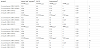
When used alone, the MIC range of LCME were 512 - 1024 μg/ ml, but in combination with AMB, CSP, TRB, and VRC, these values reduced to 64 μg/ml, 64 - 128 μg/ml, 64 μg/ml, and 64 - 256 μg/ml, respectively. Compared to the single use, the MICs of antifungal agents were also decreased in the combination tests. The previously determined MIC ranges of AMB, CSP, TRB, and VRC were 8 - 128 μg/ml, 32 - 64 μg/ml, 128 μg/ml and 8 - 64 μg/ml, respectively [14]. In combination with LCME, the MIC ranges of AMB, CSP, TRB, and VRC were decreased to 0.25 - 4 μg/ml, 0.125 - 16 μg/ml, 4 - 8 μg/ml, and 0.125 - 4 μg/ml, respectively (Table 3). Compared to the single use, the MIC values of antifungal drugs in the combination tests could be decreased to their achievable therapeutic plasma concentrations in several cases [18-21].
4. Conclusions
This is the first study investigating the activity of cysteine derivatives against Scedosporium species and the first report of the in vitro antifungal effect of LCME and its synergistic interactions with conventional antifungal agents against Scedosporium isolates. According to our results, LCME alone or in combination with other drugs might be useful for the treatment of human Scedosporium infections. However, further studies are required to investigate the antifungal mechanism of LCME and its pharmacokinetic and pharmacodynamic properties.
Competing Interests
The authors declare that they have no competing interests.
Author Contributions
László Galgóczy, Tamás Papp, Palanisamy Manikandan, and Csaba Vágvölgyi took part in the experimental design, the evaluation of the results and the final preparation of the manuscript. The single and combination tests for the fungal isolates were carried out by László Galgóczy and Mónika Homa.
Acknowledgments
L.G. holds a Lise Meitner-Position (M 1776-B20) from the Austrian Science Fund (FWF). T.P. is a grantee of J. Bolyai Scientific Scholarship of the Hungarian Academy of Sciences. The study was supported by the European Union co-financed by the European Social Fund (TÁMOP-4.2.2.B-15/1/KONV-2015-0006). This work was also funded by the grant LP2016-8/2016.