


1. Introduction
Inferior mesenteric vein (IMV) thrombosis occurs far less often than thrombosis of the superior mesenteric vein [1,2]. The IMV drains blood from the descending colon, sigmoid colon, and part of the rectum. Clinical symptoms of IMV thrombosis are mainly characterized by abdominal pain and the mortality rate remains high [1,2]. Radiologists should be familiar with computed tomographic (CT) features of IMV thrombosis in evaluating high-risk patients, therefore, optimizing medical management. We presented a rare case of a 55-year-old woman, who was early detected with CT scan, of having laparoscopy-associated IMV thrombosis.
2. Case Report
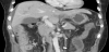
A 55-year-old woman suffered from abdominal pain for two days. She had had a 5-hour laparoscopic left hemicolectomy for sigmoid colon cancer two weeks ago. She did not receive any post op anticoagulation therapy aside from Aspirin (Bokey 100 mg/cap QD) which she has been taking for her previous stroke, other medication include amlodipine (Norvasc 5mg/tab QD) for hypertension. Physical examination revealed tachycardia (110 beats/min). Leukocytosis (15,120/uL) was noted. CT scan revealed a thrombus of the IMV (Figure 1), without extension to the portal vein, and a focally distended small bowel with wall thickening. The patient was managed conservatively in hospital with bowel rest, aggressive hydration, and prophylactic antibiotic medication. Her condition improved gradually without further symptoms and she was discharged after two weeks.
3. Discussion
Mesenteric venous thrombosis is an uncommon entity that causes mesenteric ischemia, a potentially lethal disorder with various causes. It accounts for 5 to 15% of cases of acute mesenteric ischemia and involves the superior mesenteric vein in 95% of cases [2]. Mesenteric ischemia solely due to IMV has rarely been reported [1,2]. Laparoscopy-associated IMV thrombosis is even more rare and has been identified as a complication of laparoscopic cholecystectomy, laparoscopic Nissen fundoplication, and laparoscopic Roux-en-Y gastric bypass but not laparoscopic colectomy, until now [3]. Various factors are associated with mesenteric venous thrombosis, including systemic conditions such as cancer, portal hypertension, inflammatory disease, postoperative states or trauma, morbid obesity, protein C or S deficiency, rectal vascular malformations, increased intraabdominal pressure during laparoscopic surgery leading to hindrance of splanchnic blood flow, and a hypercoagulable state [4,5].
A systematic review by James et al. [3] revealed 18 cases of portomesenteric venous thrombosis after laparoscopic surgery excluding splenectomy. Out of these, 7 cases were after Roux-en-Y gastric bypass, 5 cases were after Nissen fundoplication, 3 cases were after partial colectomy, two cases were after cholecystectomy, and 1 case was after appendectomy. Locoregional factors particular to laparoscopic procedures may contribute to the development of PVT (portal vein thrombosis). Insufflation of the abdomen and increased intra-abdominal pressure led to decreased mesenteric and portal venous flow via direct pressure–induced compression. Estimates of this decrease in venous flow vary from 35% to 84% [3]. However, most studies find a dose-dependent relationship between insufflation pressures and venous stasis. Transperitoneal diffusion of carbon dioxide into the circulation can cause hypercapnia, which in turn has been implicated in decreasing splanchnic blood flow related to mesenteric vasoconstriction [3]. Another possible explanation is that a prolonged reverse Trendelenburg position (such as may be necessaryfor various laparoscopic procedures) may exacerbate laparoscopy- associated portal venous stasis. In addition, intraoperative surgical manipulation may damage the splanchnic endothelium and lead to local thrombus formation that may then propagate throughout the portal venous system.
Nonspecific abdominal pain is the most commonly encountered symptom. The hallmark of mesenteric ischemia is abdominal pain that is out of proportion to the physical findings [2,3]. Given the rarity of this entity and the nonspecific presentation, the diagnosis is challenging. Advanced imaging techniques, such as conventional CT or CT angiography, have played a key role in rapid and accurate diagnosis.
In this case, only the thrombosis in the IMV with small bowel distension was apparent. The IMV does not drain the small bowel and in our case the sigmoid colon has been surgically removed together with the upper rectum. Therefore, the small bowel edema was common coexist CT findings, similar to previous reports [1].
History of previous stroke and hypertension, the presence of sigmoid colon carcinoma, increase intra- abdominal pressure from laparoscopic surgery, postoperative status of the patient and the surgical clips for hemostasis might have disrupted local splanchnic blood flow and may have contributed to the thrombotic event. Given that the disease can be fatal, prompt and aggressive management is essential. The lack of standard terminology used for reporting this complication is a common concern among surgeons. Some advocate the term “laparoscopy-associated mesenteric vascular complications” [4]. Accurate description of this uncommon complication is important for better understanding and management of this disease. Physicians should be aware of this rare complication following laparoscopic surgery.
Competing Interests
The authors have no competing interests with the work presented in this manuscript.