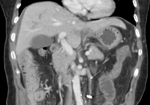
Figure 1: Coronal contrast-enhanced CT scan shows a thrombus formation (white arrow) in the inferior mesenteric vein and adjacent small bowel distension, without extension to the portal vein. Surgical clips (black arrow) placed in the mesentery are shown.