


1. Introduction
The dorsal scapular nerve (DSN) is a motor nerve that primarily originates from the fifth cervical spinal nerve root in the brachial plexus [1-6]. Occasionally, in addition to C5, the DSN may also receive contributions from C4 [7-10]. The DSN arises within the posterior cervical triangle deep to the prevertebral fascia [11] and typically pierces the middle scalene muscle where it travels posteriorly between the posterior scalene and the serratus posterior superior muscles to provide motor innervation to the levator scapulae, rhomboid minor, and rhomboid major muscles. Collectively, all three of these muscles act to elevate and retract the scapula [12-17].
Several anatomical studies in the primary literature have indicated the variability of the DSN in terms of its spinal root origins and muscular innervations. For example, Shilal et al. (2015) reported that the DSN not only receive contributions from C5 and C6 but also communicated with branches from the long thoracic nerve [18]. Similarly, Ballestero's and Ramirez's study reported that nearly 48% of the DSNs branched from C5 while nearly 30% shared a trunk with the long thoracic nerve [19]. A recent cadaveric study by Nguyen et al. (2016) found that approximately 70% of the DSN originated from C5 while 22% arose from C4 and 8% branched from C6 [13]. Chen et al. (1995) also reported that in addition to C5, the DSN received variable contributions throughout C4-T1 [20]. In addition, there are varying reports regarding the muscular innervations of the DSN. For example, a case study in Japan reported that the DSN innervated the serratus posterior superior muscle [21]. In a study by Frank et al. [22], they reported that the DSN innervated the levator scapulae muscle in only 11 out of 35 neck specimens. Similarly, Nguyen et al.'s study also found that 48% of the DSN supplied the levator scapulae muscle only whereas 52% of the nerve supplied the levator scapulae as well as the rhomboid major and minor muscles.
DSN syndrome is characterized by general symptoms of sharp, dull, or aching pain along the medial border of the scapula that radiates to the lateral surface of the arm and forearm [23]. Patients also report dysfunction of their shoulders as well as pain in their neck and back region [20]. DSN syndrome is often caused by the entrapment or impingement of this nerve at the middle scalene muscle, because the DSN often pierces this muscle [13,24-26]. However, because the DSN lacks sensory branches, the entrapment of this nerve is often overlooked during clinical diagnosis of back and interscapular pain [17,24]. In addition, the variability in the anatomy of the DSN in terms of its spinal root origins and muscular innervations may also be another factor in which DSN impingement is frequently missed [13]. Occupations that require overhead work, such as painters and electricians, make these particular individuals more susceptible to DSN impingement [17]. There are also documented injuries of the DSN amongst athletes such as volleyball and basketball players, judo, and body builders [24,27-29]. For example, along with injury to the suprascapular nerve, the DSN was also injured in two sibling volleyball players. Both siblings reported pain in their right shoulders and scapular region as well as mild winging of their right scapulas with weakness of the rhomboid muscles [30]. There are also case reports in which a lesion to or neuropathy of the DSN caused scapular winging [31-33]. For example, Akgun et al. [17] reported a 51-year-old man who damaged his DSN after lifting a heavy box overhead. As a result from this lesion, he developed right shoulder pain as well as weakness of arm abduction and winging of his right scapula.
Current treatments to help resolve patients of their pain from DSN syndrome include muscle manipulation at the scalene muscles and/ or nerve block injection [13,20,34]. According to Walther, soft tissue manipulation can be performed by passively extending the patient's neck in order to specifically stretch their middle scalene muscle of the affected side [35]. Another form of conservative treatment is directly anesthetizing the DSN. In this method, a nerve block injection that is typically guided via ultrasound, is administered in order to relieve patients of their symptoms [16,25,36,37]. Although rare, surgical intervention such as lesion of the middle scalene muscle have also been reported to relieve patients from their pain [20]. In both types of these conservative and surgical treatments, it is imperative for rehabilitation professionals to be aware of other important anatomical structures surrounding the scalene muscles of the neck such as the phrenic nerve as well as the roots and trunks of the brachial plexus in order to reduce the risk of injuring these structures.
Our previous study of the DSN investigated the relationship of this nerve as it crosses the middle scalene muscle relative to the transverse plane of the laryngeal prominence [13]. Average distances from the transverse plane of the laryngeal prominence to where the DSN entered, crossed, and exited the middle scalene muscle were reported. We used data from our previous study, then added to those anatomical data by presenting thumb interphalangeal joint (IPJ) width to approximate and predict the surface projection of the DSN. This was done relative to its site of entrapment (the middle scalene muscle) while using the transverse plane of the laryngeal prominence and the posterior border of the SCM muscle as anatomical landmarks. According to Liu et al. [38], thumb width is a convenient measurement tool commonly used by clinicians such as physical therapists to measure the distance from the location of pain to a given body landmark. Injection studies were performed to test the accuracy of using thumb IPJ width to locate the site of DSN entrapment at the middle scalene muscle.
The overall purpose of this study is to provide a convenient method for rehabilitation professionals to examine, diagnose, and treat patients with possible DSN impingement through the use of thumb IPJ width while using the transverse plane of the laryngeal prominence and the posterior border of the SCM as reference points. This method will assist clinicians in evaluating and implementing appropriate therapeutic treatments to patients who may exhibit symptoms of DSN syndrome.
2. Materials & Methods
The surface projection of the dorsal scapular nerve was examined in 10 embalmed adult cadavers (6 males and 4 females) obtained through the Willed Body Program, Center for Anatomical Sciences, at the University of North Texas Health Science Center (UNTHSC) in Fort Worth, Texas. The age of the donors span from 68 to 92 years with a mean age of 80 years. The self-reported ethnicities of the donors are Caucasian. The cadavers are individually wrapped in cotton shroudswith Maryland State Wetting agent (Hydrol Chemical Company, Yeadon, PA.) and are stored in metal tanks located in the UNTHSC Gross Anatomy Laboratory.
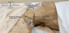
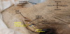
The cadavers used in this study have not been previously dissected and therefore, all skin in the neck region remained intact. The posterior border of the sternocleidomastoid (SCM) muscle was first identified and palpated. A transverse plane through the laryngeal prominence was established using a 90º-angled ruler. A grease pencil was used to outline the posterior border of the SCM muscle as well as mark the transverse plane of the laryngeal prominence to create reference points. An injection was made at approximately 2.08 cm medial from the intersection of the posterior border of the SCM and the transverse plane of the laryngeal prominence (Figure 1(a)). This value is the average distance at which the DSN exited the middle scalene muscle from the transverse plane of the laryngeal prominence as reported from our previous research [13]. In addition, 2.08 cm is equivalent to approximately one thumb IPJ width as reported from Liu et al.'s study in which average thumb IPJ width is approximately 2.0 ± 0.4 cm [38]. For injection, a polyurethane resin (PU4ii) with a proprietary blue dye was prepared following the manufacturer's instructions (vaQtec, Zürich, Switzerland). Approximately 0.1 ml of the resin dye was injected at a depth of 1 cm using a 1 ml syringe with a 22 gauge needle. The polyurethane resin was allowed to solidify for 24 hours post-injection. Dissections were then made along the posterior border of the SCM to reveal the location of the injection site as indicated by the blue dye. The distance of the dye to the DSN was measured using an electronic sliding caliper (Carrera Precision Corp.). All injections and dissections were performed on the left side of the neck region. On the right side of the neck, a previous incision was made to access vasculature for the embalming of our cadavers. Therefore, important structures such as the scalene muscles and the DSN were often damaged on that side. Dissection images were taken with a digital camera (Nikon Coolpix S6200).
3. Results
The surface projection of the DSN was investigated in 10 embalmed adult cadavers. Measurements were also taken between the site of injection and the actual location of the DSN at the midpoint of the middle scalene muscle. The results of the injection study revealed that in 5 cadavers, the resin dye was located directly at the middle scalene muscle as the DSN either pierces or crosses anteriorly to this muscle. On one cadaver, the dye was located at the anterior scalene muscle and the distance between the location of the dye to the DSN was 0.683 cm. On two cadavers, the dye was located between the anterior and middle scalene muscles. The average distances between the location of the dye and the DSN at these injections was approximately 1.40 cm. In another cadaver, the dye was between the middle and posterior scalene muscles and the distance between the location of this injection to the DSN was about 0.676 cm. On the last cadaver, the dye was found at the posterior scalene muscle and the distance between this injection site and the DSN was 0.832 cm. Figure 1 is an example of our injection study showing the blue dye at the middle scalene muscle and the DSN crosses anteriorly to this muscle. It was also observed that in relation to the middle scalene muscle, 50% of the DSN pierced this muscle whereas 40% of the DSN crossed anterior to the middle scalene muscle and 10% of the nerve traveled posterior to the middle scalene muscle.
4. Discussion
We used previous data from Nguyen et al. [13] in order to estimate the surface projection of the DSN relative to the middle scalene muscle. The average distance, 2.08 cm (± 0.96 cm), was chosen from our previous research as the distance for the injection site from the intersection of the transverse plane of the laryngeal prominence and the posterior border of the SCM muscle. This distance for the injection site was chosen for several reasons. Because 2.08 cm is the measurement at which the DSN exited the middle scalene muscle, this value is located at the most lateral border of this muscle. Therefore, important anatomical structures such as the phrenic nerve and the superior trunk of the brachial plexus would be farthest away from the injection site. This information is especially important for rehabilitation professionals in order to avoid injuring these anatomical structures during a nerve block injection. In addition, for therapists and clinicians, 2.08 cm is approximately 1 thumb IPJ width which makes this measurement clinically useful in pinpointing the surface projection of the DSN while using the reference points of the posterior border of the SCM muscle and the transverse plane of the laryngeal prominence.
The results of our investigation revealed that the surface location of the anterior, middle, and posterior scalene muscles were consistently identified when approximating the surface projection of the DSN using 1 thumb IPJ width medial to the intersection of the posterior border of the SCM muscle and transverse plane of the laryngeal prominence. Although we accurately identified the surface location of the DSN at its typical entrapment site (the middle scalene muscle) in 50% of the injections performed, the distances between the dye at other sites within the scalene muscles to the actual location of the DSN were measured. In those measurements, the average distance between the injected dye and the DSN was less than 1.0 cm which is less than half the distance of 1 thumb IJP width. Clinically, rehabilitation professionals could use these measurements as a radius to approximate the area of a circle at or very near to the DSN's position at the middle scalene muscle. This would allow professionals to treat patients with DSN syndrome by performing circular tissue manipulations within the surface projection of the middle scalene muscle.
5. Conclusion
Because the surface projection of the DSN has not been previously reported, the overall significance of this research is to provide easily identifiable reference points for clinicians to locate the nerve. Utilizing the posterior border of the SCM muscle as well as the transverse plane of the laryngeal prominence, clinicians' ability to accurately and efficiently locate the site of DSN entrapment will improve. In addition, using these reference points combined with a simple 1 thumb IPJ width measurement, this method may prove to be very useful for rehabilitation professionals to examine, diagnose, and conservatively treat patients with DSN impingement. Future studies in investigating the effectiveness of our method in a patient population along with locating the DSN via ultrasound could be beneficial in validating our proposed method.
Competing Interests
The authors declare that they have no competing interests.
Author Contributions
Vuvi Nguyen: contributed to the concept and design, data acquisition, data analysis and interpretation, and writing of manuscript. Hao (Howe) Liu: contributed to the concept and design, data interpretation, and manuscript editing Armando Rosales: contributed to data interpretation and manuscript editing Rustin Reeves: contributed to the concept and design, data analysis and interpretation, and manuscript editing for final approval
Acknowledgments
The authors thank the selfless gifts made by body donors to the Willed Body Program, Center for Anatomical Sciences at the University of North Texas Health Science Center in Fort Worth. This research would not be possible without their generosity.