


1. Introduction
Insidious Onset Mechanical Neck Pain (IOMNP) is a common cause of discomfort and disability and a growing problem in society, resulting in significant impact on health care costs [1]. Within 6 months, 54% of adults experience IOMNP, and 5% of them complain of associated disability. Furthermore, only 6.3% of people who suffered from neck pain are free from recurrence [2]. About 0.6% of the general population is exposed to disability caused by IOMNP and females are more prone to recurrent and / or chronic pain than males [3]. The IOMNP can be considered a work-related disorder in some professions, including pilots [4-7], musicians [8-9], and workers using personal computer for prolonged times [10-14].
Cervical spine supports the head and orients it in space, transmits forces on the upper limb and has close relations with the visual and vestibular function [15-18]. Given the complexity of this region and the close link between central nervous system (CNS) and muscles, several abnormalities in structure and function of the neuromuscular system may develop when pain occurs [2,15].
The aim of this review is to provide an updated overview of the changes in structure and function of the cervical neuromuscular system associated with IOMNP.
2. Methods
MEDLINE and SCOPUS databases were searched by a reviewer, using the following search string: "neck pain" AND (muscle OR "motor control" OR behavior OR function OR endurance OR "isometric contraction") NOT (radiculopathy OR surgery OR "specific neck pain"). Other studies were included after having analyzed the references of the selected papers. Search was limited to the papers published after 01-01-2003, in English or Italian languages. Observational studies on changes in structure and / or function of the cervical neuromuscular system associated with IOMNP were included. Studies on subjects with specific neck pain (e.g. spondylolisthesis, neuromuscular disorders, radiculopathy) or previous surgery were excluded. Studies on neck pain treatment and on temporomandibular joint dysfunction were also excluded.
3. Results
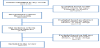
After deleting doubles items, the search yielded 1441 results. 1201 articles were excluded by reading titles, 174 by reading abstracts and 16 by reading full texts. Finally, this review included 15 papers concerning changes in structure of neck neuromuscular system, 19 on changes in pattern of neuromuscular activation, and 16 dealing with deficit in neuromuscular function associated to IOMNP (Figure 1). Results were organized and presented by different topics. Changes in cervical neuromuscular system structure (histology and metabolism, cross sectional area and ratio of size, muscle fatty infiltration, trigger points generation, and changes in muscular activation pattern) and changes in neuro-muscular function (reduced maximal voluntary contraction, altered proprioception, coordination and complex functions).
3.1 Changes in cervical neuromuscular system structure
3.1.1 Histology and metabolism
Intra- and extra-cellular environment of the neck myofascial system showed altered homeostasis in subjects with IOMNP. Analyzing fascial interstices and sliding systems with different methods, some interesting although still preliminary data emerged.
The blood flow afferent to the upper trapezius was found decreased in subjects with IOMNP, with a consequent reduction in oxygen concentration and exchanges at capillary level [19]. This reduction has been interpreted by the authors as a consequence of sympathetic nervous system overactivation. Takiguchi et al. [20] found similar results in subjects with pain induced by intra-muscular injection of hypertonic solution in trapezius muscle. The rapid occurrence of these changes suggests a direct involvement of the sympathetic nervous system proximally to the peripheral β-2 adrenergic receptor, whose activity remains unchanged.
In upper trapezius of subjects with IOMNP an increase of paininducing substances as serotonin, glutamate, interleukins and E2 prostaglandin was found [21-22]. In upper trapezius of chronic patients lower concentration of N-acetyl-ethanolamine compared to the rest of the body was also observed [23]. Evidence about the alteration of muscle temperature was on the contrary few and contradictory [24].
Important anomalies were also observed within the muscle fibers. Three case reports published by Green et al. [25] appeared as very interesting to understand intracellular biochemical changes of the myo-fascial syndrome associated with IOMNP. These authors found a decrease of O2, decrease in phospho-kinase activity, decrease [up to 45%] of Ca reuptake within the sarco-plasmatic reticulum, and disruption of ATPhasic Na-K.
From a functional point of view, already in the 90s several studies highlighted the transformation of muscle fibers from Type I to Type II. This type of fiber processing seemed directly related to the increase in EMG fatigue in sub-maximal tasks [26].controller around the wrist and the battery and computer in the backpack weighing 3.5kg [26].
3.1.2 Cross Sectional Area [CSA] and Ratio of Size [ROS]
The Ratio of Size [ROS] indicates the relationship between muscle cross-sectional area [CSA] and Body Mass Index [CSA / BMI = Ratio of Size]. The ROS is reduced in subjects with IOMNP, in particular on the affected side, if the pain is unilateral. This phenomenon is shown at all cervical levels regardless of the area in which symptoms are referred. Association was also found among loss of lordosis, pain intensity and disability. These findings come from studies on multifidus, longus collis and cervical semispinalis muscles [27-28]. The reduction of ROS seems to be due to the decrease of muscle thickness in anterior-posterior direction, not in medial-lateral one [28].
3.1.3 Muscle Fatty Infiltration [MFI]
MFI, widely reported in literature, is the infiltration of fat in cervical muscles, in particular on type I fibers and deep muscles. Deep extensors, predominantly tonic, are more interested compared to deep flexors, which have an equitable distribution of slow and fast fibers. This phenomenon is often associated with increased CSA and its pathogenesis is still unclear [29].
ElIiott [29] showed by NMR that MFI happens in patients with WAD. In fact, subjects with IOMNP have data of MFI comparable to the healthy population, whereas patients with WAD are significantly affected (p <0.001).
3.1.4 Trigger Points [TP] generation
Myofascial syndrome is a painful condition that affects muscles and provokes local and / or referred pain. This syndrome is characterized by an increased presence of active and latent TP compared to healthy tissue, increased extrinsic stifness, tenderness, and change in motor control of the affected area. The evidence indicates a direct correlation between neck pain (of any origin) and increased presence of TP [30].
In IOMNP the increased presence of TP is localized only in superficial muscles (in particular sternocleidomastoid, scalenus anterior, upper trapezius, levator scapulae, and some sub-occipital muscles), unlike the WAD, in which deep muscles such as the cervical semi-spinalis are also affected.
3.1.5 Changes in muscular activation pattern
This review summarizes the results of the studies which investigated activation pattern through instrumental diagnostic methods such as electromyography (EMG), Real-time Ultra-Sound (RUS) and Muscular Functional Magnetic Resonance (mfNMR).
These studies were always carried out on various cervical levels, however significant difference between different segments has never been found. Literature reports four main alterations that characterize the pathological pattern of cervical muscles activation in patients with IOMNP.
Increased activity of superficial muscles and decreased of the deep ones: These phenomena have been observed in tasks that require only neck stabilization or minimum contribution of superficial muscles, such as in cranio-cervical flexion or upper limb movements [31-33]. The decreased activity of deep muscles was also found in isometric contractions in different directions, which physiologically require the action of both superficial and deep muscles [34-35].
The correlation between pain, even though artificially induced, and this altered pattern was demonstrated by Cagnie et colleagues [36-37]. They showed through mfNMR an immediate shift towards altered physiological contraction after injection of hypertonic solution, in tasks as head flexion and neck extension. A direct correlation between activation of the superficial muscles during cranio-cervical flexion and pain intensity was also demonstrated in patients with IOMNP. However, there was no evidence of correlation between muscular activation and duration of pain and disability.
Although these data confirm the important role of pain in generating the pathological pattern, other contributing factors were found [31]. Some studies [12-13] have shown that, in patients whose pain appears after a certain time of activity (such as violinists or those who use keyboard), the pattern of alteration typical of IOMNP have begun before the onset of pain. Furthermore, pathological pattern and kinematic alterations always seem to be associated.
Non-specific muscle activity: This phenomenon was shown through two altered behaviors of the cervical muscles: coactivation of agonists and antagonists and non-specific direction. In particular, hyper-activity of the splenius capitis during cervical flexion and hyper-activity of the sternocleidomastoid during cervical extension has been recorded in patients with IOMNP [1,38]. This agonists and antagonists co-activation seemed related to pain, disability and decrease in muscle strength [1].
It was also observed that agonists and antagonists co-activation during low-intensity tasks was greater in flexion and lateral flexion towards the dominant limb, compared to the extension and lateral flexion towards the not dominant limb [39]. An association between directional aspecificity (and the consequent appearance of parasitic movements) and IOMNP was also been demonstrated.
Falla et al. [40] recorded EMG activity of the sternocleidomastoid muscle in subjects with IOMNP during multi-directional isometric contractions. Compared to the control group, patients had both inability to modulate the recruitment of motor units [MU] in relation to the direction of contraction, and an always maximal activation of the sternocleidomastoid.
Inability to relax after muscle activation: The EMG signal was found altered in patients with IOMNP compared to controls: it was reduced during an isometric contraction and increased during the resting phase of [1,40]. Wytrazek et al. [41] also found a correlation between amount of EMG signal alteration and presence of active TP. It was also observed a reduced Flexor Relaxation Phenomena (FRP) in subjects with IOMNP, indicating an inability to relax the extensor muscles when maximal neck flexion occurs [42].
Deficit in deep flexors anticipatory timing, especially during upper limb movements: This alteration was observed in subjects with neck pain for at least a year, with no significant differences between IOMNP and WAD [43]. Similar alteration was observed during neck movements in subjects whose pain was induced by hypertonic solution injection [36-37]. The immediate effect of the pain on the anticipatory timing suggest a prompt change in CNS activity.
Falla et al. [44] found a direct correlation between lack of anticipation (in upper limb movements and in cranio-cervical flexion) and pain intensity in patients with chronic IOMNP. No correlation instead appeared with duration of symptoms or disability. In addition, the alteration of activation pattern and the functional reduction were more impaired in tasks of low intensity (25% of Maximal Voluntary Contraction - MVC) with respect to medium intensity (50% MVC) and high intensity (100% MVC) tasks [32].
Other changes in neuro-muscular system behavior: In patients complained of acute and chronic IOMNP or Low Back Pain (LBP), an increased evoking reflexes foot area was found, without significant correlation between duration of symptoms and dimension of this area [45].
Finally, Richter et al. [46] found a lower Reaction Time [RT] during Hand Mentaly Rotation [HMR] tasks in patients with WAD, compared to healthy subjects and patients with IOMNP. The decrease of RT was proportional to the duration of pain. The group of patients with IOMNP instead presented values similar to those of healthy subjects.
3.2 Changes in neuro-muscular function
3.2.1 Maximal Voluntary Contration [MVC]
In patients complained of neck pain for more than a year, the MVC in flexion, extension, rotation and lateral flexion appeared significantly lower than in healthy subjects. In particular, MVC was reduced by an average of 22.6% in flexion, 33.2% in extension, and 32.2% in lateral flexion movements [1,47]. A moderate correlation between pain experienced during the test, fear of movement and reduced performance was also highlighted [35].
Finally, an MVC reduction of the lower trapezius compared with the contralateral during upper limb movements in subjects with IOMNP was also observed.
3.2.2 Proprioception and Coordination
There was conflicting evidence regarding the presence of alteration in proprioception, measured with the Joint Position Error [JPE] in patients with WAD compared with healthy subjects. The repositioning errors were generally small, namely from 2° to 5°. Instead, patients with IOMNP seemed to have no alteration in proprioception compared to healthy subjects [48-49].
Nevertheless, using measures different from JPE some authors observed deficit in proprioception in patients with neck pain [2,50]. Woodhouse & Vasseljen [48] found deficit in combined movements of the head and lower accuracy in patients with WAD and IOMNP compared to healthy controls. No difference was instead found between controls and other groups using JPE. Moreover, Malmstrom et al. [51] showed that repositioning errors increased with artificially induced pain in healthy subjects.
3.2.3 Muscular endurance
In subjects with IOMNP a deficit of muscle strength in both deep and superficial muscles was found [52-53]. The fatigue was similar in different movement directions and did not appear affected by symptomatic vertebral level.
The endurance impairment was even greater for low intensity contractions. Specifically for contractions at 50% of MVC the maximum time was reduced by 27%, whereas for tasks at 25% of MVC the reduction was 35% compared with healthy subjects [54]. Furthermore, in case of unilateral IOMNP, fatigue was superior in muscles ipsilateral to the pain [55]. This deficiency, ipsilateral to the pain, was also found in upper trapezius during upper limb movements [56].
The endurance deficit was related to the duration of pain in patients with IOMNP from less than five years. After this time period, the fatigue remained, but its magnitude was no longer correlated to the duration of pain [57].
Parazza et al. [58] found a significant correlation between deep cervical flexors and extensors endurance and among endurance, pain intensity and disability. No relationship was instead found between reduced endurance and stage of pain (acute, subacute or chronic).
4. Discussion
The results of this review suggests that pain, whether it be artificially produced or due to IOMNP, causes an alteration of cervical region function and several anomalies in behavior and structure of neuromuscular system.
Pain causes an immediate reduction in blood flow to the upper trapezius with a consequent reduction in O2 concentration and exchanges at capillary level [19-20]. This modification on the vascular system has been interpreted as orthosympathetic response to pain and it is related with O2 reduced presence and reduction of ATP activity [25].
The finding of algogenic substances (serotonin, glutamate, E2 prostaglandin, and interleukins) in cell interstices, and the reduced presence of N-acetyl-stenolammine (endogenous lipids implicated in regulation of inflammation and nociception) suggest that these biochemical alterations may provoke nociceptors sensitization. Homeostasis is altered to a greater extent in areas clinically known as TP, especially in active TP. It seems that TP do not constitute separates entities, but rather areas in which such anomalies are particularly relevant [62]. The association between the amount of EMG signal alterations and presence of TP emphasizes the close relationship between changes in structure (which are exemplified by TP) and changes in neuro-muscular function.
The reduced availability of oxygen, caused by the reduction of blood supply, can promote the shift from type I to type II fibers in affected muscles. These changes could explain the increased muscular fatigue and weakness in submaximal tasks, typical of patients with IOMNP [26]. The disuse syndrome, linked to pain and kinesiophobia, could be responsible for the decreased ratio of size in patients with IOMNP. However, muscle fatty infiltration has been demonstrated to be exclusive of patients with WAD [29,63,64].
One of the functional consequences of the cross sectional area reduction seems to be the maximal voluntary contraction deficit: this affects people with IOMNP from at least one year and concerns movements in different directions [1,47]. The amount of this reduction appears very different among various studies, due to several factors, including heterogeneity between individuals and mechanisms of inhibition. In fact, pain is perceived during strength test by more than 65% of patients and it appears moderately correlate with fear of movement and reduced performance [35].
Changes in structure and function of the neuromuscular system could probably be triggered from a response of the CNS to painful stimulus, at least in the first phase. In addition to the sympathetic system, it is reasonable to speculate also the involvement of pyramidal and extrapyramidal system. It could explain the immediate shift from a physiological pattern of muscle activation to an altered one, in response to pain induced by intramuscular injection of hypertonic solution [36-37].
The pathological pattern has been observed in neck and in upper limb movements, that both require stabilization by deep cervical muscles. The first responses to pain include widespread spinal cord hyper-excitability, which is demonstrated by the increased evoking reflexes foot area in patients with acute IOMNP [45].
The abnormal movement patterns could be not only an effect of pain, but also a factor contributing to pain itself. This hypothesis is supported by the finding that in some subgroups of subjects undergoing continuing upper limbs tasks, pain appears after a certain time of activity and alterations in pattern of movement are observed before pain appears [12-13,65].
In chronic patients, a direct correlation between pain and impairment is no longer detected. For example, after 5 years, the duration of pain is not related to the amount of endurance reduction, while it is in acute and subacute phase [57]. After few years of pain probably the importance of other factors such as disuse and kinesiofobia increases [34].
We emphasize two important findings from this review of the current literature.
- The amount of agonist-antagonist co-activation is directly related to the disability [31]. This anomalous behavior is interpreted as an effort of the nervous system to maintain stability at segmental and regional level, involving mobilizing muscles. Unfortunately, this co-contraction neither ensures stability, nor favors movement or function.
- The extent of the deep cervical flexors endurance deficit is closely related to that of the deep extensors and is associated to pain intensity and disability. No association was found instead from endurance reduction and pain stage [58].
An interesting finding that emerges from the literature is the absence of altered proprioception [measured by JPE] in subjects with IOMNP [48-49]. Despite this, many authors reported clinical experience of relevant alterations of movement precision. A possible interpretive hypothesis is that the coordination and precision deficitIn JPE test proprioception is involved, but it is not particularly impaired in subjects with IOMNP. In contrast, in subjects with WAD from more than a year proprioceptive information is altered and visual channel is favored, with the consequence of improving the performance in head mentaly rotation tasks [46].
By a clinical point of view, we stress the absence of any correlation between neck symptomatic level and structural, functional or behavioral alterations, which involves the entire region with some differences only as regards the more affected left or right side [34].
Finally, we point up adverse effects of IOMNP on complex functions not so closely related to the cervical region: breathing dysfunction [59], shoulder girdle disorders [60,66], and alterations in gait kinematics [61].
5. Conclusion
The relationship between IOMNP and structural or functional changes of cervical neuromuscular system are now acquired evidence and there is a widespread consensus about the fact that these alterations contribute to the onset and maintenance of neck pain and promote recurrence. The changes that occur rapidly (as the modification of the vascular system or the shift toward a pattern of pathological activation) are most likely mediated by the CNS, as more directly related to pain. Other impairment instead which appear after a certain period of time, such as fatigue, may be more attributable to changes intrinsic to the muscle, being influenced by other factors such as disuse and kinesiophobia.
There is still a lack of literature on the alterations of complex functions (such as breathing, walking and activities of the upper limb).
Future studies are suggested to investigate the sequence and the causal relationships between the different impairment and to investigate the effectiveness and type of therapeutic exercise or manual therapy can positively influence changes in muscle structure and function.
Competing Interests
The authors declare that they have no competing interests.
Author Contributions
All authors contributed substantially to conception and design, acquisition and analysis of data and interpretation of results.