


1. Case Report
A 9-year-old female child had a known case of Turner Syndrome with congenital heart disease associated with repaired coarctation of the aorta and ventricular septal defect VSD. Moreover, the patient was known to be short stature and receiving Somatotropin hormone treatment. The patient was admitted to the emergency room complaining of high fever (>38.0°C for 48 hours), non-bilious vomiting repeated three times, right foot swelling associated with hotness, redness, and tenderness.
The patient also complained of pain in the swelling area. There was no associated history of any other medical symptoms like cough, shortness of breath, nor any changes of taste or smell sensations. Besides, there was no history of any trauma, insect bites, nor scratches at the swelling area.
General examination of the patient revealed an alert, conscious, oriented to time, place, and persons with no difficulty breathing. Initially, the temperature was 39.2 °C, oxygen saturation of 98%, heart rate of 138 beats per minute, respiratory rate of 22 breaths per minute, and blood pressure of 116/78 mmHg. On local examination, there was external deformity and swelling of the right foot; furthermore, edema and erythema with severe tenderness were present at the plantar and dorsum of the right foot. Dorsalis pedis artery pulse was felt, no lymph nodes enlargement.
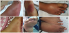
During the COVID-19 pandemic and due to high fever the patient presented with, a routine nasopharyngeal swab, and blood panel tests were done, which indeed showed positive results PCR for SARS-CoV-2, the total leukocyte count was 12,800 cell/mm³ with a differential neutrophil count of 89.9%, lymphopenia of 760 cell/mm³, platelets 313,000 cell/mm³, and ESR of 73 first hour mm/hr. CRP 15.2, LDH 209 IU/ L (normal reference: 60 -170 IU/L), Ferritin 74.1 ng/ ml (normal reference: 10-200 ng/ml), Troponin-I 0.8 pg/ml (normal reference: 0-350pg/ml), plain chest X-ray was normal, right footplain X-ray showed soft tissue swellings. Ultrasound with doppler for the right foot showed normal blood flow without any fluid collection or compression of deep fascia, ECHO with doppler showed a provisional normal pericardium, and coronaries. Blood culture was sent, and Empirical therapy of intravenous ceftriaxone and clindamycin, Zinc sulfate, 25-hydroxy vitamin D, and Enoxaparin sodium LMWH were started (Figure 1a, Figure 1b, & Figure 1c).
The patient's manifestations improved after 5 days of continuous intravenous antibiotics, and blood culture was negative with no organism detected. Moreover, another nasopharyngeal swab was sent for PCR which showed negative results for the SARS-CoV-2. Total leukocyte count decreased to 5,240 cell/mm³, neutrophils count 52.4% with a return of lymphocyte count to 2000 cell/mm³, CRP 5.4, ESR 55. Anti Xa 0.3 U/L, and the repeated Anti Xa was 0.1 U/L as Enoxaparin sodium was discounted (Figure 1 D, E, & G).
2. Introduction
Turner syndrome is characterized by monosomy of X chromosome, a structurally abnormal X-chromosome or mosaicism with a peculiar phenotype, the main manifestations are short stature, lymphoedema, sexual infantilism, cardiovascular, renal, skeletal, and a wide spectrum of skin abnormalities particularly melanocytic naevi and skin appendage abnormalities like pterygium colli. However, there are frequent ichthyotic changes, café‐au‐lait spots and halo naevi. Hypertrichosis, nail changes and keloid formation are frequently seen [1].
Congenital lymphoedema of the hands and feet is a common feature in Turner syndrome [2].
Lymphoedema is a chronic, debilitating and incurable condition. It is characterized by the accumulation of lymph and other elements (commonly proteins) in the interstitial spaces. This is due to a failure of the lymph-conducting system. A compromised lymphatic system results in lymphatic insufficiency, with swelling and fluid retention in one or more limbs or body segment. Turner syndrome is one of the genetic causes of primary lymphoedema [3].
Cellulitis is a rapidly spreading serious bacterial infection of the skin and subcutaneous fat that is typically acute in onset, with diffuse erythema that spreads over the course of hours to days. It appears as red, tender, hot swelling usually in the foot, face, or arms [4]. Occasionally, there is inflamed regional lymph nodes or associated lymphangitis. It is usually preceded by a history of trauma, scratch, or insect bite in the area of infection occasionally associated with abnormal immune function [5].
The novel second coronavirus created a pandemic. It was associated with two main characters: The highly contagious nature and the intercontinental spread with a high impact on the global economy and public health [6]. This novel beta coronavirus was named severe acute respiratory syndrome coronavirus 2 (SARS-CoV-2), and the disease it causes was called coronavirus disease 2019 (COVID-19) [7].
Human beings could be infected by SARS-CoV-2 through the respiratory tract due to expiratory respiratory droplets or contact with contaminated objects by the virus. During the primary stage of this epidemic, the infection spread to the community mainly through person-to-person contact. At this point, transmission almost occurred among adults. After this initial stage, in mid-January 2020, the disease started to be transmitted within family members, spreading to children and older adults [8].
The first pediatric case infected with SARS-CoV-2 in a 10-year-old child occurred about a week after a trip to the city of Wuhan. He was asymptomatic but had ground-glass opacities on the chest computed tomography CT [9].
Infected symptomatic children showed a predominance of fever (22.2-100%), cough (11.1-75%), associated with gastrointestinal symptoms, including nausea, vomiting, diarrhea, abdominal pain (8.8- 57.1%), and abnormalities in lung imaging (40-100%). Occasionally there are increased serum creatine kinase MB isoenzyme (CK-MB) levels, C-reactive protein (CRP), and procalcitonin. Less frequent laboratory findings included leukocytosis, leukopenia, lymphopenia, lymphocytosis, neutropenia, and increased transaminase levels and erythrocyte sedimentation rate (ESR). The need for oxygen was low (2.3-28.6%), except in the study by Sun et al., who described only severe cases in children [10].
Kawasaki disease (KD) pathophysiology is complex and postulated to be secondary to overactive innate and adaptive immune system in genetically predisposed individuals [11-18].
Kawasaki disease (KD) criteria for diagnosis Table 1.
The Case Definition for Multisystem Inflammatory Syndrome in Children (MIS-C) according to the Centers for Disease Control and Prevention (CDC) [19], are listed in Table 2.
3. Discussion
Wu et al., described a child aged 2 years and 10 months with conjunctivitis, eyelid dermatitis, normal chest CT, increased LDH and CK-MB, lymphocytosis, and neutropenia, with positive nasopharyngeal swab PCR for SARS-CoV-2. Seizures have also been reported in children infected by SARS-CoV-2 [20].
A papulovesicular eruption all over the trunk sparing other body sites was reported in 8-year-old girl, she had 6-day history of cough, PCR-test was positive with mild thrombocytopenia [21].
A 10-year-old boy with positive PCR-swab was admitted to a hospital in United Kingdom with tender plantar papules, macules, and petechiae allover his legs, few days later an annual patch appeared. Papules were also noted in his axilla [22].
In Iran a 12-month-old infant with SARS-CoV-2 PCR positive swab had Erythema-multiform-like eruption with targetoid lesions on his trunk, and extremities, along with acral erythema, and pyrexia [23].
To diagnose Kawasaki disease (KD) patient should have fever for ≥ 5 days with ≥ 4 “Other features”, or evidence of cardiac involvement like (coronary aneurysm, myo/pericarditis, or pericardial effusion) [24].
The current SARS-CoV-2 pandemic is associated with a higher incidence of severe cases of Kawasaki-like disease in children from different regions of the world. The pathophysiology of these presentations is still unknown but may be related to the cytokine storm detected in severe manifestations of COVID-19 in adults [25].
Although the features of Multisystem Inflammatory Syndrome in Children (MIS-C) are like those of KD, but there are few different distinct entities. Only less than 25% of MIS-C are fulfilling KD diagnostic criteria. Patients with MIS-C are usually older than those with KD, have higher white blood cell count, neutrophil count, and CRP, as well as more profound lymphopenia and anemia. They also tended to have lower platelet counts, higher fibrinogen levels, and greater elevation of troponin. Alanine aminotransferase levels and D-dimer levels are similar between MIS-C and KD cases [26].
The current case was <21 years presented with fever, laboratory evidence of inflammation, and evidence of clinically severe illness that required hospitalization, with multisystem (>2) organ involvement (hematologic, gastrointestinal, and dermatologic). There were fever(>38.0°C for > 24 hours), repeated vomiting, right foot swelling associated with hotness, redness, tenderness, and no lymph nodes enlargement, there is hematologic inflammatory evidence and SARSCoV- 2 positive PCR test.
Co-existing of COVID-19 and Cellulitis could be attributed to vasculitis associated with MIS-C, lymphedema that associate Turner syndrome, part of skin manifestations of COVID-19, or incidental.
4. Limitations
This case study had some limitations. First, it was based on one case study with significant symptoms during COVID-19 pandemic at a period before and during the development of the case definition. Investigations and management were individualized by center and patient rather than following WHO standardized protocol.
Second, there is no diagnostic test for MIS-C or KD, so it is not possible to clear cut a definite diagnosis of the cellulites with such diseases.
5. Conclusion
A predictive correlation between SARS-CoV-2 infection and cellulitis was referred in the current case study , the co-existence of COVID-19 and Cellulitis could be attributed to vasculitis associated with MIS-C, lymphedema that associate Turner syndrome, part of skin manifestations of COVID-19, or incidental. A further research is needed to establish a definite diagnosis, relation, and pathogenesis.
Competing Interests
The authors declare that they have no competing interests.
Author Contributions
AbdElShakoor KI conceived the ideas, collected &analyzed the data and led the writing.
Alzahrani MS managed the case and collected the clinical data.