


1. Introduction
Cancer of the uterine cervix is the third most common gynaecologic cancer diagnosis and cause of death among gynaecologic cancers in the United States [1] . Cervical cancer has lower incidence and mortality rates than uterine corpus and ovarian cancer, as well as many other cancer sites. These rankings are similar to global estimates for other developed countries [2] . Women with locally advanced cervical cancer (stage IB2 to IVA) have a higher rate of recurrence and worse survival than those with early-stage disease (stage IA to IB1). After surgery alone, the rate of relapse is at least 30 percent, and five-year survival rates range from 80 percent for stage IB disease to 30 percent for stage III disease [3,4]. Exenteration has been used for the last 6 decades, mainly to treat cancers of the lower and middle female genital tract in the irradiated pelvis. New ablative techniques based on developmentally derived surgical anatomy termed laterally extended endopelvic resection (LEER) aim to increase the curative resection rate, even of tumours extending to and fixed to the pelvic side wall5. The aim of this report is to describe the case of a patient with locally advanced cervix cancer who is treated by LEER and other reconstructive techniques in order to improve her quality of life. conservative approach.
2. Case Description
A 24-year-old woman presenting moderately differentiated squamous carcinoma in biopsy. Physical exam reveals a thickened cervix with bleeding ulcers and a mass of 20 mm is observed in posterior cervical lip by ultrasounds.
Analytically only emphasize a raised SCC levels. The extension study with MRI show a diffuse thickening of the cervix of approximately 60x50x45 mm infiltrating upper third of the vagina, uterine inferior third and regions 6 cm from the anus. As well as 3 left iliac nodes and other bilateral inguinal ones.
After studies she is staged as locally advanced cervix and paraaortic lymphadenectomy retroperitoneal staging is done to determine the level of radiation, being negative the nodes studied. She begins chemotherapy (weekly CDDP) concomitant with external radiation therapy (TD: 50.4Gy) and complete gynaecological brachytherapy PDR (HR 30Gy), which ended with complete response.
Eight months later a RMI control is done, where a mass of 4 cm appears affecting the rectum and right parametrium. PET-CT is applied, and it excludes extrapelvic disease so the patient is proposed to LEER as a part of the total pelvic exenteration , intraoperative radiation therapy with electrons (IORT), urinary diversion (Indiana type) with both vaginal and rectum reconstruction.
3. Description of Surgical Procedure
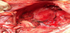
LEER (Laterally extended endopelvic resection) was based on total mesorectal excision, total mesometrial resection, and total mesovesical resection. The inclusion of pelvic side wall and floor muscles, such as the obturator internus, pubococcygeus, iliococcygeus, coccygeus muscles, and eventually of the internal iliac vessel system (Figure 1).
IORT was delivered on the right parametrial bed (infiltrated in MRI) and left parametrium (surgical infiltration), with pelvic nerves protection. The administered dose was 12.5 Gy (90% isodose pattern). The applicator used was 5 cm, 45 degree bevel and energy of 6 MeV in both locations.
The continent urinary diversion (Indiana pouch) was made after IORT. Firstly ureters were mobilized, doubly clipped. The clips were left on the proximal and end of the ureters until it was time to connect them to the colon reservoir. Then the entire right colon, hepatic flexure, proximal transverse colon, cecum and terminal ileum were mobilized. The mesentery of the right colon was transilluminated to determine the blood supply, and the sites for division of the transverse colon. About 24-30 cm of colon and 12-15 cm of ileum were needed, typically this would include the cecum, ascending colon and 4-6 cm of transverse colon. The mesenteries were incised through the avascular areas down to the posterior peritoneum.
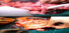
After cleaning the isolated colon segment, it was opened along the antimesenteric border (through the taenia libera) excising the appendix. To get the continence mechanism the ileum was plicated over a Foley catheter with 3-0 silk sutures from the ileocecal valve to the level of the linear stapler. The redundant bowel was then removed with the linear stapler. The plication might be continued along the length of the ileal segment inverting the staple line. After this, the opened colon was folded upon itself and stay sutures placed at the four corners. The lateral edge was closed with an inverting, running stitch of absorbable suture. Then the reservoir was positioned in its approximate final location to estimate the degree of ureteral mobilization required for an anastomosis without tension, kinking or angulation (Figure 2). For the ureteral anastomoses the ileal segment was pulled through the stomal tunnel to approximate the final position of the reservoir. The ureters had already been mobilized and brought into position under the ascending mesocolon (Figure 3). One was on either side of the suture line in the posterior wall of the reservoir. The stomal apertura was made through the abdominal wall in the right lower quadrant. The last step was to complete the reservoir, for this, a large Malecot catheter was brought into the pouch and secured with an absorbable purse string suture. The ileal segment was pulled through the stomal aperture and the reservoir positioned against the anterior and lateral abdominal wall.
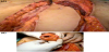
To build a neovagina a rectus abdomenis myocutaneous flap was used. An elliptical cutaneous paddle 12 cm in diameter that had a vertical axis centered over the upper half of the rectus abdominis muscle. The paddle extended from the costal margin to just below the umbilicus; its lateral borders extended to within 2 cm of palpable muscle, to enhance the viability of the lateral paddle margins (Figure 3A). A vertical incision was made through skin, subcutaneous tissue, and anterior rectus fascia. The rectus muscle was divided using electrocautery, with care taken to ligate the superior epigastric vessels on the posterior aspect of the muscle. To prevent separation of muscle from the anterior rectus fascia, which might devitalize the paddle, the muscle was fixed to the edge of the overlying fascia and skin with interrupted sutures. The remaining muscle was carefully dissected free from the posterior rectus fascia to the pubic symphysis, with care taken to preserve the inferior epigastric vessels. The paddle was tubularized by folding the paddle in half (Figure 3B), the lateral edge and apex were closed with interrupted suture, and the neovagina was rotated into the pelvis without tension and sutured to the vulvar introit. A resection of the sigmoid colon and the rectum were performed during the exenteration and a terminal colostomy was made to ensure digestive diversion.
Two drains, one in Douglas and the other in subcutaneous, were left. As intraoperative incidences the patient had tachycardia and a transfusion of two packed red blood cells was required as well as a unit of frozen plasma.
4. Postoperative Follow-Up
After surgery, the patient was transferred to the intensive care unit for 8 days and 17 days after surgical procedure was discharged from hospital.
In subsequent outpatient controls, patient has several episodes of UTIs which are treated with oral antibiotics. The clinical and image controls performed alternately every 3 months after surgery, were normal one year after surgery and a 10 cm length stenotic vagina was found at clinical examination with a good the quality of life.
5. Discussion
Growing evidence indicates that local tumor spread is not random but is confined to permissive tissue compartments defined by embryonic development [6] .
Compartmental borders are natural barriers for neoplastic propagation. Locally recurrent tumours are not only advanced in malignant progression but grow in a tissue landscape that may have been altered through the previous anti-cancer treatment. Particularly after surgical therapy compartimental borders may have been damaged and substituted by scar tissue, which appears to be associated with a loss of their barrier function and to forward neoplastic transgression.
Therefore, local tumor relapses are often multicompartmental even at small sizes. According to the theory of compartmental tumor spread, advanced and recurrent tumors may require multicompartmental resection for local tumor control [5] .
Laterally extended endopelvic resection (LEER) is aimed to resect en bloc multiple visceral compartments in the female pelvis within intact borders for local control of advanced and recurrent malignancies of the lower female genital tract [7,8].
In our patient, after completing initial treatment with chemotherapy and radiation therapy, no signs of radiological disease appeared, nevertheless, the cervical cancer mass relapsed months later. So it was decided to perform a LEER procedure following this theory.
In surgical treatment of the pelvic recurrent disease the free margins of the specimen is mandatory. IORT can provide an additional dose of irradiation on a well-defined area, which is considered a high risk of local recurrence.
The continent reservoir (Indiana Pouch) was created in a classic manner, although in literature is described that appendix can provide a convenient continence mechanism, in our patient is not applicable as she had pelvic radiation because of the associated atrophy and fibrosis of the appendix. We preferred this type of continent reservoir instead of the Miami pouch because of the high rate of complications of the last one in case of prior pelvic radiation [9] .
Gracilis myocutaneous flaps have been used most commonly to create a neovagina in young people. However, gracilis flaps are associated with a high failure rate (11–37%) due to vascular compromise; leave unsightly scars on the medial thighs; are frequently too large and bulky to use when the perineal body and anus are preserved [10,11]. In our patient we prefered a vertical myocutaneous flap to create the neovagina and one year after the intervention, the patient has none of these complications and it has achieved an increase in its quality of life, although a long-term monitoring is necessary to assess late complications.
6. Conclusion
LEER has a significant potential role to rescue selected patients with locally advance or recurrent gynecologic cancers, including those with pelvic side wall disease. The best results in this type of salvage surgery are when free margins are obtained. The use of IORT as a part of the procedure ensures the microscopic treatment of the margin without any secondary effect.
In young patients the reconstructive procedures improve the quality of life after the treatment. The continent reservoir employed in our patient is well tolerated and the needless plastic bag used in another type of urinary diversion also improves quality of life. In the same way the neovagina created with a rectus adominis flap decreases the feeling of mutilation by the patient.
In conclusion in selected experienced teams in tertiary centers and in selected patients this salvage surgery can be the last opportunity of cure of these patients.
Competing Interests
The authors declare that they have no competing interests.