


1. Introduction
Brain natriuretic peptide (BNP) is a biomarker with multiple functions, for example, diuresis, natriuresis, and arterial vasodilation, leading to reduced activities of the renin-angiotensin-aldosterone system and the sympathetic nervous system [1]. These biological actions of BNP are considered to be protective against impaired hemodynamics in patients with heart failure (HF). Measurement of BNP level is useful in the diagnosis of acute decompensated HF in patients with dyspnea in emergency departments [2,3]. In addition, the level of BNP is predictive of in-hospital mortality [4] and shortterm prognosis after discharge (less than 1 year) [5-7] in hospitalized patients with acute decompensated HF. However, the predictive value of BNP level on long-term prognosis is still unclear in patients with acute decompensated HF.
Pre-pro-BNP, known as the precursor of biologically active BNP, is synthesized in the ventricular myocardium and released into the blood circulation in response to ventricular wall stress [8,9]. According to Laplace's law, the wall stress in a sphere increases in proportion to the radius and decreases in inverse proportion to the wall thickness [10,11]. Thus, left ventricular (LV) morphology is considered to be an important determinant of LV wall stress or BNP secretion, but little data is available regarding the effect of LV morphology on the prognostic value of BNP levels in patients with HF. Thus, the objective of this study was to assess the long-term prognostic value of BNP levels, in relation to LV morphology, in patients hospitalized with acute decompensated HF.
2. Methods
2.1 Study population
The present study consisted of consecutive patients who were hospitalized at Matsushita Memorial Hospital for acute decompensated HF from April 2000 to August 2002. Inclusion criteria were ≥18 years of age and no history of myocardial infarction, stroke, major surgery, or active malignancy diagnosed within the last 3 months. A diagnosis of acute decompensated HF was defined as new-onset or progressive worsening of symptoms with pulmonary and/or systemic congestion definitely or probably due to elevated ventricular filling pressure [12]. All patients were treated in accordance with the standard guidelines for HF [13].
A total of 117 patients with acute decompensated HF met our inclusion criteria. Of these, 27 patients were excluded from the analyses: 18 patients who died during the hospital stay, 3 who were later transferred to other hospitals, 3 who underwent or were scheduled to undergo surgical intervention (coronary intervention in 2 and aortic valve replacement in 1), and 3 with chronic hemodialysis. Ultimately, this prospective study consisted of 90 patients who were hospitalized for acute decompensated HF and discharged after treatment (38 women; 34 to 87 years of age, mean age 69 years). Informed consent for this study was obtained from all patients.
2.2 BNP
Plasma levels of BNP were measured before discharge with stable hemodynamics using previously described methods [14]. In brief, ethylenediaminetetra-acetic acid-treated blood was processed and frozen at -20 degrees Celsius after centrifugal separation within 20 minutes. The assays were conducted using a commercially available kit (Shionoria BNP, Shionogi & Co., Ltd., Osaka, Japan). The analytical range and normal reference range of the assay were 4.0 to 2,000.0 pg/ ml and <18.4 pg/ml, respectively.
2.3 LV morphology
All patients underwent transthoracic echocardiography in a hemodynamically stable condition by a standard method before discharge [15]. In brief, LV end-diastolic internal dimension, LV endsystolic internal dimension, ventricular septal wall thickness, and LV posterior wall thickness were measured on 2-dimensional or M-mode echocardiograms using a commercially available ultrasound system (Sonos 5500; Philips Medical Systems, Andover, Massachusetts, USA). LV end-diastolic volume was determined as (LV end-diastolic internal dimension3 × 7)/(2.4 + LV end-diastolic internal dimension); and LV end-systolic volume was determined as (LV end-systolic internal dimension3 × 7)/(2.4 + LV end-systolic internal dimension) [16]. LV mass was determined according to the equation of 1.04 × [(LV end-diastolic internal dimension + LV posterior wall thickness + ventricular septal wall thickness)3 - LV end-diastolic internal dimension3] × 0.8 + 0.6 [17]. LV end-diastolic volume, LV endsystolic volume, and LV mass were indexed by body surface area defined as weight0.425 (kg) × height0.725 (cm) × 0.007184 [18].
2.4 Outcome
All patients were prospectively followed after discharge for the composite endpoint of cardiac events defined as cardiac death or rehospitalization due to decompensated HF. Cardiac death was defined as death from progressive HF, myocardial infarction, arrhythmia, or sudden death. Sudden death was defined as a witnessed death within 1 hour after the onset of symptoms or an unwitnessed death in a patient known to have been alive and functioning normally 24 hours before. Rehospitalization due to decompensated HF was defined as the development of pulmonary edema requiring intravenous treatments (inotropic agents, vasodilators, or diuretics), mechanical ventilation, or circulatory support. All patients were classified into 3 groups according to their outcomes: survivors without cardiac events, survivors with cardiac events, and nonsurvivors, at the times of 1 year, 5 years, and 10 years after discharge. During the time course, the status of patients could change from survivors without cardiac events to survivors with cardiac events or nonsurvivors, or from survivors with cardiac events to nonsurvivors. Patients were excluded if they died due to noncardiac causes during the follow-up period, for example, a patient who died of cancer 6 years after discharge was included in the assessment at 1 year and 5 years after discharge, and excluded from the assessment at 10 years. Patient information was obtained from available medical records and interviews with patients or their family physicians.
2.5 Statistics
Baseline characteristics among survivors without cardiac events, survivors with cardiac events, and nonsurvivors at 10 years after discharge were compared by Chi-square test for categorical variables and 1-way analysis of variance for continuous variables. The levels of BNP among the 3 groups at 1 year, 5 years, and 10 years after discharge were compared using the Kruskal-Wallis test because of possibly skewed distributions. Spearman’s rank correlation coefficient was used to examine correlations between BNP levels and LV morphologyincluding LV end-diastolic volume index, LV endsystolic volume index, and LV mass index. A 2-sided P value <0.05 was considered statistically significant. All statistical analyses were performed using SPSS version 11.0J (SPSS Inc., Chicago, Illinois, USA).
3. Results
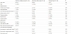
Follow-up data were available in 89 patients at 1 year after discharge, 87 patients at 5 years, and 84 at 10 years; 1 patient was lost to follow-up within a year after discharge; 5 patients died due to noncardiac causes during the follow-up period (cancer in 2, aspiration pneumonia in 1, infectious disease of unknown origin in 1, and unknown in 1). At 1 year after discharge, 6 patients had died and 26 patients had survived with cardiac events. At 5 years after discharge, 20 patients had died and 26 had survived with cardiac events. At 10 years after discharge, 28 patients had died and 24 had survived with cardiac events. No cardiac events occurred after discharge in the remaining 32 patients during the follow-up period of up to 10 years. Baseline characteristics at enrollment, as stratified by cardiac events at the time of 10 years after discharge, are shown in Table 1. Cardiac events were associated with older age, lower estimated glomerular filtration rate, and higher levels of BNP. LV morphology including LV end-diastolic volume index, LV end-systolic volume index, and LV mass index did not differ significantly among survivors without cardiac events, survivors with cardiac events, and nonsurvivors. The levels of BNP were not correlated with LV end-diastolic volume indexes (r = 0.15, p = 0.16), LV end-systolic volume indexes (r = 0.16, p = 0.15), or LV mass indexes (r = 0.08, p = 0.45).
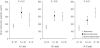
As shown in Figure 1, at 1 year after discharge, BNP levels before discharge were highest in survivors with cardiac events (357 ± 71 pg/ ml; range, 3 to 1500 pg/ml), followed by nonsurvivors (240 ± 107 pg/ ml; range, 7 to 669 pg/ml), and lowest in survivors without cardiac events (177 ± 23 pg/ml; range, 13 to 840 pg/ml). At 5 years after discharge, BNP levels were equally higher in survivors with cardiac events (307 ± 66 pg/ml; range, 7 to 1500 pg/ml) and in nonsurvivors (300 ± 63 pg/ml; range, 3 to 1010 pg/ml) than in survivors without cardiac events (157 ± 24 pg/ml; range, 13 to 690 pg/ml). At 10 years after discharge, BNP levels were higher according to the severity of the outcome: 357 ± 65 pg/ml (range, 3 to 1500 pg/ml) in nonsurvivors, 222 ± 43 pg/ml (range, 7 to 840 pg/ml) in survivors with cardiac events, and 145 ± 25 pg/ml (range, 13 to 690 pg/ml) in survivors without cardiac events.
4. Discussion
The present study was conducted to clarify the long-term prognostic value of BNP levels for cardiac events, in relation to LV morphology, in patients hospitalized with acute decompensated HF. The main finding was that long-term prognosis was associated with BNP levels before discharge, but not with LV morphology, in patients with acute decompensated HF. Another important finding was that the prognostic value of BNP levels on the severity of outcome (i.e., death versus rehospitalization versus no event) varied depending on the time after discharge.
Dynamic changes in BNP levels have been observed during hospital stay, but these levels before discharge seem to be the most reliable in predicting the short-term prognosis in patients with acute decompensated HF. Daily measurement of BNP levels in 72 patients hospitalized with acute decompensated HF [5] showed that these levels before discharge were a strong predictor of death or rehospitalization 30 days after discharge. Similarly, in another study consisting of 105 patients hospitalized with acute decompensated HF [7], serial measurements of BNP levels showed that these levels before discharge were reliable in predicting the risk of death or rehospitalization 6 months after discharge. We found that BNP levels before discharge were also predictive of long-term outcome, up to 10 years, in patients hospitalized with acute decompensated HF.
In the present study, no association of LV morphology with BNP levels before discharge was found regarding long-term outcomes in patients hospitalized with acute decompensated HF, although LV morphology is considered to be closely associated with LV wall stress or BNP secretion [8-11]. The insufficient adaptation of LV morphology to rapidly worsening conditions in patients with acute decompensated HF may be a cause of the discrepancy between BNP levels and LV morphology in our cohort. This speculation may be, in part, supported by the fact that the diagnostic accuracy of BNP levels for HF differs between patients with acute decompensated HF and patients with chronic HF [19], implying that our results cannot be applied to the assessment of patients with chronic HF.
In the present study, higher levels of BNP before discharge were associated with early rehospitalization due to decompensated HF (i.e., within 1 year after discharge) and late cardiac death (i.e., 10 years after discharge) in patients hospitalized with acute decompensated HF. Interestingly, at 5 years after discharge, the levels of BNP were equally high in patients who were rehospitalized due to decompensated HF and patients who had cardiac death. These findings may be useful in selecting patients who will benefit from additional treatment to avoid future cardiac events. Further interventional studies are needed to examine whether BNP-guided treatment, considering the time after discharge, could improve outcomes in patients hospitalized with acute decompensated HF.
5. Study Limitations
The present study was conducted in a single center and our cohort was highly selected; the present findings may not be extrapolated generally to patients with acute decompensated HF.
6. Conclusion
Long-term prognosis was associated with BNP levels before discharge, but not with LV morphology, in patients hospitalized with acute decompensated HF. The prognostic value of BNP levels on the severity of outcome varied depending on the time after discharge.
Competing Interests
The authors declare that they have no competing interests.
Author Contributions
All the authors substantially contributed to the study conception and design as well as the acquisition and interpretation of the data and drafting the manuscript.