


1. Introduction
Cancer is one of the most feared of all human diseases, and the need to develop high-efficacy and low-toxicity cancer drugs is an important field that is being actively studied. Consecutively occurring impairments to DNA result in activation of several oncogenes, including Myc. In contrast, tumor suppressor genes are inactivated, which affects the DNA repair system and disrupts normal apoptosis regulation pathways [1]. Myc is a transcriptional regulator with a basic helix-loop-helix leucine zipper (bHLH-ZIP) domain that becomes functional after dimerization with its obligate partner protein, Max [2]. The coiled-coil structure of the Myc-Max heterodimer has a palindromic E-box sequence 5'-CACGTG-3' that recruits the DNA [3]. Myc interacts with target gene promoters to stimulate or suppress transcriptional activity [4]. Overexpression of Myc is involved in transformation processes, including proliferation, apoptosis, differentiation, and metabolism [5]; however, dysregulation of Myc is involved in the vast majority of human cancers such as lung, pancreatic, and colorectal cancer in addition to leukemia and lymphomas [6]. Because of its multiple functions, there has been concern that targeting Myc-Max/DNA interactions for drug development would result in undesirable side effects. However, studies of the dominantnegative Myc mutant, omomyc, have suggested that pharmacological inhibition of Myc results in gentle and reversible effects on normal, quick-proliferating tissues [7,8]. Together, these studies suggest that Myc inhibition by direct disruption of Myc-Max/DNA or subsidiary inhibition of Myc using BET bromodomain inhibitors [9-11] could be a feasible restorative method and is on the cutting edge of new and focused anticancer drug development. The favored methodology to identify possible Myc inhibitors has been to measure obstruction of Myc-Max dimerization; however, protein-protein interactions (PPIs) within the large surface area (~3,200 Å2) and many binding pockets that have not yet been identified are primary hindrances [3]. Despite these difficulties, an absolute amino acid substitution may completely disturb the dimerization of Myc with Max [3]. Here, we provide evidence for the principle that a high-affinity ligand attached to the interactional surface may provide additional disruption of Myc-Max dimerization.
2. Myc Inhibitors
Procownik et al. applied a yeast two-hybrid assay systemtoa library of 10,000 chemicals to discover 10058-F4, a rhodanine scaffoldcontaining small molecule that is a potent Myc-Max inhibitor [12]. An HL60 human promyelocytic leukemia cell line over-expressing Myc was then used to test the efficacy of 10058-F4. Myc-Max/DNA binding was interrupted by 10058-F4, with an IC50 value of 49 μM. Considering the strong protein-protein interactions involved in Myc- Max/DNA binding, including strong hydrogen bonding, electrostatic interactions, and π–π interactions, we suggest that these results are remarkable. From the perspective of medicinal chemists, small molecules contain many positive attributes, including the ability to substitute several functional groups on the scaffold. In the work by Procownik et al., several substituents on the 4-ethylphenyl moiety of 10058-F4 were introduced, including 4-chlorophenyl; cyclohexyl; 3,4-dihydroxyphenyl; and 4-isopropylphenyl moieties; however, substituted compounds have not improved the activity of 10058-F4. Fixing a 4-ethylphenyl group on the left position of 10058-F4 and then modifying the right side of the rhodanine core did not significantly improve activity. In a follow-up modification, N-methylpiperidinyl functionalization of the rhodanine NH group followed by substitution with an isopropylphenyl ring resulted in the introduction of 28RHNCN- 1 and inhibition of Myc binding to DNA that is equivalent to 10058-F4 in an in vitro system. However, the inhibitory activity increased 2-fold in HL60 whole cells, with an IC50 = 29 μM. Both 28RH-NCN-1 and 10058-F4 demonstrated concentration-dependent inhibition of Myc-Max heterodimer formation in HL60 cells that was associated with a decrease in cell proliferation [13].
Work done in the laboratory of Metalloet al. from Georgetown University elucidated the interruption mode of 10058-F4 and 10074- G5 to their target proteins Myc-Max using NMR dynamic structures. 10058-F4 binds Myc402-412 sequences, whereas 10074-G5 binds Myc363-381 sequences [14]. In addition, the nitro group and furazan core of 10074-G5 could interact with the positively charged amino acid residues, Arg 372 and Arg 362, while the biphenyl group has π-π interactions with a hydrophobic region composed of Phe375, Ile381, and the side chain methylene moiety of Arg 378. The discovery of two free tying sites, each comprising over 10 residues inside a bHLH-ZIP domain that is 84 amino acids in length, suggests that sites capable of specific small-molecule binding are universal in intrinsically disordered proteins [14] (Table 1).
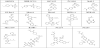
Work by Fletcher et al. identified the Myc inhibitor pharmacophore, 10074-G5, through a structure-activity relationship (SAR) study. 10074-G5 is composed of three distinct moieties, including a nitro functional group on the top subunit, a benzofurazan core, and a 2-aminobiphenyl group. Reduction of the nitro group to an amine and introduction of several functional groups on the NH2-nitrogen atom resulted in loss of inhibitory activity. Modification of the biphenyl moiety to phenyl, hydroxyphenyl, cyclohexyl, methoxyphenyl, bromophenyl, and piperidinyl resulted in no or weak inhibition activity; however, introducing a carboxylic acid at the para-position on the phenyl ring (compound JY-3-094) increased the potency to five times of that observed for the lead compound, with an IC50 of 33 μM for disruption of Myc-Max heterodimers [15]. In a follow-up paper to their early work, Fletcher et al. introduced ester functionality to the para-position of JY-3-094, which has poor cell permeability. The prodrug molecule JY-3-094 and its derivatives warrant further development of small-molecule Myc-Max inhibitors [16]. Recently, Fletcher et al. disclosed the active and stable (half-life in cell > 17 h) Myc-Max inhibitor 3jc48-3 [17], which has an additional hydrophobic group on the phenyl ring of the prodrug molecule JY-3-094. Treatment of HL60 and Daudi cells with 3jc48-3 repressed proliferation with single-digit micromolar IC50 values in a way that corresponded to the intracellular interruption of Myc-Max heterodimers. In addition, Fletcher et al. developed a novel and direct Myc inhibitor, JKY-2- 169, that is distinctly different from their previous compounds. This new molecule contains a bis-benzamide core via synthetic α-helix mimetics. JKY-2-169 perturbs Myc-Max heterodimer binding to approved E-box DNA sequences without producing protein-protein dissociation.The resulting inhibition of Myc in proliferating cells, both in vitro and in vivo, leads to cell cycle arrest and apoptosis. Compared with previous Myc inhibitors, JKY-2-169 does not disrupt Myc-Max heterodimers in cells, but specifically inhibits a Myc-dependent reporter. Consequently, JKY-2-169 binds an α-helical region of Myc and decreases the ability of theMyc-Max heterodimer to bind DNA without provoking the dissociation of Myc-Max heterodimers. Several bis-benzamide derivatives exhibited a 2-fold or greater selectivity for Myc-Max heterodimers over Max-Max homodimers with IC50 values of 5.6 μM [18].
Vogt et al. isolated and validated the first nonpeptidic Myc-Max inhibitors from an in-house peptidomimetics library using an ELISA and electrophoretic mobility-shift assay (EMSA). Among these inhibitors, IIA6B17 containing an isoindoline scaffold antagonized the Myc-Max/DNA ternary complex with an ELISA IC50 value of 125 μM and EMSA IC50 value of 40 μM [19]. Moreover, IIA6B17 hindered Myc-induced oncogenic transformation of chicken embryo fibroblast (CEF) cells (IC50 = 20 μM). In addition, the lead molecules developed by Vogt et al. was discovered to inhibit c-Jun, inferring that their mechanisms might involve the bHLH-ZIP domain that is found in many transcription factors. It is possible that these compounds restrain the oncogenic activity of Myc indirectly by stabilizing Myc- Max homodimers and thereby diminishing the concentration of Max accessible for heterodimerization by Myc [20]. Together with Boger et al., Vogt and colleagues substituted the isoindoline-5,6-dicarboxylate scaffold of IIA6B17 to a pyrrolidine-3,4-dicarboxylate group (labeled mycmycin-1 and mycmycin-2), resulting in inhibition of Myc-induced oncogenic transformation in CEF cells. The second generation of Myc inhibitors designed by this group is 10-fold more potent than the parent compound, IIA6B17. Notably, mycmycin-1 and mycmycin-2 do not inhibit the oncogenic transcription factor c-Jun.
Janda, Vogt, and colleagues developed a compound library based on a reciprocal hydrophobic and planar naphthalene scaffold. Forty small molecules were screened from 285 compounds through a fluorescent resonance energy transfer screening assay. Among the 40 screened molecules, 4 effectively inhibited Myc-Max/DNA binding, including NY2276 and NY2267, with IC50 values in the range of 17–36 μM. NY2276 has an IC50 value of 17 μM in an in vitro assay; however, this compound has weak antagonistic effects on Mycinduced oncogenic transformation in cell culture [21]. Recently, these researchers identified compound KJ-Pyr-9 in a Krőhnke pyridine library using a fluorescent polarization screen. KJ-Pyr-9 is not acutely toxic at concentrations as high as 10 mg/kg and penetrates the bloodbrain barrier. In addition, KJ-Pyr-9 occurs at higher concentrations in brain tissue than in the blood after 4 h. Furthermore, nude mice treated daily with 10 mg/kg KJ-Pyr-9 by i.p. injection for one month expressed MDA-MB-231 cells. Tumor volume in the KJ-Pyr-9-treated mice did not notably increase and body weights were unchanged [22].
Berg et al. utilized a fluorescent polarization assay to screen a library of 17,000 chemicals in order to discover a Myc inhibitor, Mycro 1, which disrupts the binding between Myc-Max and DNA with an IC50 value of 72 μM. Mycro 1 is selective for Myc-Max heterodimers over c-Jun–c-Fos heterodimers and inhibits proliferation of a diverse number of cell lines, including the breast cancer cell line MCF-7. In a recent progress report, Berg et al. reported the discovery of Mycro3, a pyrazolo[1,5-a]pyrimidine scaffold that inhibits both Myc-Max dimerization and DNA-binding with good selectivity [23].
Henriksson et al. identified Myc pathway response agents (MYRA), which are small molecules that inhibit Myc-induced cellular transformation[24]. Interestingly, MYRA-A and MYRA-B have different inhibitory effects on the DNA-binding activity of Myc- Max. MYRA-A hampers the DNA binding of Myc-Max and Mnt- Max heterodimers in a dose-dependent manner, but has no effect on DNA binding of the E-box-binding transcription factor, USF. In contrast, MYRA-B showed no effects at a concentration of 400 μM, as determined by EMSA. MYRA-A might conceivably discriminate the Myc protein from other E-box-binding proteins; however, MYRA-B potentially acts through effects on variant E-box promoters by interacting with other transcriptional factors that bind Myc, by interfering with adaptor proteins, or by an indirect mechanism [24].
3. Challenge
Our understanding of mechanisms involved in direct interruption of Myc-Max heterodimerization or Myc-Max/DNA complexation using small molecules has been advancing rapidly over the past several years. Several research groups have identified small molecules that have good inhibitory effects on the Myc-Max/DNA complex, in both in vitro and in vivo models. Experimental data suggest that small molecules have the potential to be advanced into therapeutic agents for customized cancer treatment. However, we still need to overcome significant barriers in order to obtain highly specific and active anticancer drugs. The development of small molecules using rational drug design, along with functionalization to interrupt the hot spot between Myc-Max and Myc-Max/DNA could result in the development of effective therapeutic drugs to treat cancers.
Competing Interests
The authors declare that they have no competing interests.