


1. Introduction
Klebsiella pneumoniae have been documented as the common factor for cryptogenic liver abscess in Asia Pacific [1,2]. Similar findings in North America and Europe have also been reported [3-5]. Several virulent factors of K. pneumoniae were discovered and these factors are almost related to specific capsular polysaccharides (CPS) [2,6-8]. Accumulating reports have indicated that the capsule is essential to the virulence of Klebsiella species [9-11]. Up to now, the capsular antigen have been classified into 77 serotypes , and serotype K1 and K2 capsular antigen were found to comprise virulence in mouse peritonitis model, whereas isolates of serotypes other than K1 or K2 were with little or no virulence [5,12].
Outer membrane porins (OMP) are proteins that cross a cellular membrane and contribute to the diffusion of molecules. Many studies have showed OMPs play important roles in antimicrobial resistance [13,14]. Previously, we have demonstrated the virulent effects of OMPK 36 by an animal study [15]. In this report, the results of animal lethality study showed that the virulence of OmpK36 mutant was reduced about 100 fold compared to the wild-type strain NVT- 1002, which was isolated from a patient with liver abscess. However, the participation of OmpK36 in K. pneumoniae induced liver abscess formation and the toxicity to cells are still unclear. In this study, we examined the role of OmpK36 in K. pneumoniae induced pathogenesis by animal model and the inflammatory cytokines were also detected. In addition, the direct toxic effects of OmpK36 and whole bacteria were detected by the cell model.
2. Materials and Methods
2.1 Mice liver abscess study
Klebsiella pneumoniae isolates NVT-1002 and ΔOmpK36 were obtained as previously [15]. The adult BALB/c mice were injected intraperitoneally with 103 cfu of K. pneumoniae NVT-1002 or ΔOmpK36-NVT-1002 in 0.1 mL phosphate buffered saline. Mice were sacrificed with CO2 after 1-3 days bacterial injections, the livers were removed, fixed with paraformaldehyde and embedded in paraffin. Histological sections were observed after hematoxylin–eosin (H&E) staining.
2.2 Kinetic of inflammatory cytokines production in liver and serum
Liver was removed for tissue section and supernatant of each sample was collected for cytokines production. Cardiac blood samples were collected under aseptic conditions. Blood samples were allowed to colt at 40°C and then centrifuged at 15000 rpm for 3 min. Serum samples were preserved at -80°C until measurement of the cytokine. Cytokine concentrations in liver and serum were measured with a mice cytokine 10-plex antibody bead kit (R&D systems, Minneapolis, MN) according to the manufacturer's instructions. Tumor necrosis factor (TNF)-α, interleukin (IL)-1β, IL-6 and IL-10 levels were determined. The supernatants from homogenized liver were then withdrawn at the indicated times, and dilutions were assayed in triplicate for each independent experiment. All samples were tested in triplicate. The minimal detectable protein concentration was 10 pg/ml.
2.3 Construction of pET30a-OmpK36
The DNA fragment of OmpK36 was amplified by primer pairs OmpK36F (5’-GGGAATTCCATATGCACCATCATCATCATCATA TGAAAGTTAAAGTACTG-3’) and OmpK36R (5’-CCGCTCGAG GAACTGGTAAACCAGGCC-3’) from a clinical isolate NVT-1002. The amplified fragments were digested with restriction enzymes and cloned into the expression region of plasmid pET30a (Novagen). The pET30a-OmpK36 was used to express a 6His-OmpK36 protein as described below.
2.4 Recombinant OmpK36 protein expression, purification and detoxification
The pET30a-OmpK36 was transformed into BL-21 (DE3) competent cells. An overnight cell culture (3 mL) was grown at 37°C in the presence of 50 mg/L kanamycin. After transfer of the cell culture to 300 ml of Luria-Bertani medium, the cell suspension was allowed to reach an OD600 of 0.7–0.9 before addition of IPTG (1 mM). Cells were grown for 4 h at 37°C and then centrifuged at 9000g for 30 min. Whole-cell lysates were prepared by sonication (Sonics Vibracell sonicator, 25% amplitude, pulsed 1 sec “on” and 2 sec “off” for a total of 5 min of “on” time, at 4 sec) in phosphate buffered saline with 6 M urea. The soluble protein fraction was then mixed with 8 mL of Ni2+ resins (Amersham) to capture the His-OmpK36 recombinant protein. The Ni2+ resins was then washed with 40 mL of wash buffer (10 mM imidazole, 6 M urea, 10% glycerol in phosphate buffered saline (PBS)) and eluted by wash buffer with 40 mM imidazole. The flowthrough was collected and dialyzed by PBS with 3M urea and 10% glycerol for 4 hr and then by PBS with 10% glycerol overnight. The endotoxin was removed by Pierce Detoxi-Gel endotoxin removal gel (Thermo Scientific, Rockford, IL) as described in user’s manual. Expression of the His-OmpK36 protein was determined Western blot and the final protein concentration was measured by Bradford assay (Bio-Rad).
2.5 Culture of hepatoma HepG2 cells
HepG2 cells was cultured in Eagle's Minimum Essential Medium supplemented with 10% fetal bovine serum (FBS), incubated at 37°C, 5% CO2 in a humidified incubator. The culture medium will be changed every day. Cells were grown to confluence in 25 cm2 tissue flasks. Subsequently, the cells was trypsinized with trypsin/ EDTA solution in phosphate-buffered saline (PBS), centrifuged and resuspended in fresh media. Before the experiment, cells were seeded onto the variant tissue culture dishes overnight.
2.6 Cell viability assay
Cell viability was detected by MTS assay, Cell titer 96®AQueus (Promega). In brief, 2×10⁴ cells were seeded on to 48 well plates in 200 μl serum free media per well and incubated for 24 hrs. After variant treatments included of a multiplicity of infection (MOI) of 60 whole bacterial infection or OmpK36 recombinant protein, cells were washed with PBS 3 times, 20 μl MTS reagents were added to each well in the final volume of 100 μl and placed in incubator for 2 hours. The optical density was measured at 490 nm using a plate reader.
2.7 Statistical analysis
Student’s t test was used for statistical analysis. Data represented as means ± standard deviations. P values of less than 0.05 was considered statistically significant.
3. Results
3.1 Contribution of OmpK36 the K. pneumoniae induced liver abscess
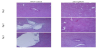
Histological sections showed that remarkable injuries in liver were seen after 24 hours of injection with wild type K. pneumoniae (NVT- 1002). More severe injuries were found at day 2 and day 3 (Figure 1). In the meanwhile, the injuries were almost abolished in mice injected with OmpK36-KO. This is suggested that OmpK36 plays an important role in K. pneumoniae induced liver abscess (Figure 1).
3.2 Rols of OmpK36 in Klebsiella pneumoniae induced cytokines expression, included of TNF-α, IL-1β, IL-6 and IL-10
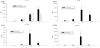
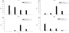
Pro- and anti-inflammatory cytokines secretion in response to bacterial infection included IL-1β, IL-6, IL-10 and TNF-α were detected by ELISA. The wild-type K. pneumoniae NVT-1002 elicited the secretions of all the cytokines both in plasma (Figure 2) and liver (Figure 3). It is notable that most of the elevated cytokines have the peaks at day 2. In contrast, the OmpK36 KO mutant only transiently enhanced IL-1βexpressions and the other cytokines were almost undetectable both in liver and serum.
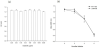
Direct cytotoxic effects of OmpK36 recombinant protein and whole bacterial infection to HepG2 hepatoma cells.
HepG2 hepatoma cells were incubated with various concentrations of OmpK36recombinant protein for 24 hours or infected with whole bacteria with a MOI of 60 for 2-6 hours. The viability of HepG2 cells were then detected by MTS. Results showed that no significant difference was detected in the toxicity to HepG2 cells (Figure 4a). Similar results were found in the toxicity of whole bacterial infection, the cell viability was not altered in the ΔOmpK36 group compared to the NVT-1002 group (Figure 4b).
4. Discussion
Liver abscesses caused by K. pneumoniae were first reported in Taiwan [1,16]. The extra-hepatic complications spread by blood stream such as endophthalmitis, fasciitis and meningitis have been reported [1,17-19]. The syndrome was subsequently reported in Asia and America [20,21]. To date, the syndrome is emerging world wildly [22]. Several virulent factors of K. pneumoniae were described, including lipopolysaccharide, capsular polysaccharide, siderophore, resistance to phagocytosis and serum killing [23]. K. pneumoniae strains expressing capsular type K1 or K2 antigen are especially virulent. [7,24]. Previously, we first demonstrated that OmpK36 is a virulent factor of K. pneumoniae. However, the involvement of this porin in K. penumoniae induced liver abscess progression is still unclear. The wild-type NVT-1002 used in this study is a serotype K1 clinical isolate which was isolated from patient with liver abscess. Injection with NVT-1002 intraperitoneally can induce severe hepatic injuries in experimental mice (Figure 1). On the contrary, the injuries were almost abolished in OmpK36 KO mutant group (Figure 1). Although this model cannot simulate the disease completely, it is still suggested that OmpK36 plays an important role in the pathogenesis of K. pneumoniae induced liver abscess.
Endogenous TNF-αis associated with severity of hemorrhagic and thrombotic lesions in organs, especially in the liver and kidney [25,26]. IL-6 and IL-10 have the potential to inhibit TNF-α and IL-β expression from macrophage and peripheral monocytes [27,28]. IL-6 also has the anti-inflammatory potential by activation of transcription-3 protein in hepatocytes [27]. The balance of the inflammatory and anti-inflammatory cytokines may determine the tissue damage and mortality [29]. A previous report has been demonstrated that high IL-10 / TNF-α ratio is associated with the mortality [30]. In this study, the serum TNF-α, IL-6 and IL-10 were elevated and have the peaks at day 2 after injection of wild type NVT- 1002 (Figure 2). Similar results were found in hepatic IL-6 and IL- 10 (Fig. 3). At the time point, sever injuries were observed in tissue sections (Figure 1). It is possible that the expressions of cytokines are associated with the pathogenesis of liver abscess. In the OmpK36 deficient group, both serum and hepatic IL-1βwere elevated, the other cytokines were almost undetectable (Figure 2 and Figure 3). This is evident that OmpK36 participate in the K. pneumoniae induced immune responses. However, the detail cellular and molecular mechanisms need further studies.
To identify the toxic effect of OmpK36, the in vitro cell model was used in this study. The HepG2 cells were treated with various concentrations of OmpK36 and the cell viability was detected by MTS method. Unexpectedly, the viability of HepG2 cells was not affected by recombinant OmpK36 protein (Figure 4a). Results of whole bacterial toxicity to HepG2 also showed that there is no significant difference was observed (Figure 4b). This is suggested that OmpK36 has no direct toxic effect to HepG2 cells. Previous study has been demonstrated that OmpK36 contributes to the resistance to phagocytosis, serum killing and the bacterial clearance in hepatocytes [15]. OmpK36 might prolong the duration of bacterial burden subsequently enlarge the injuries in organs such as liver and kidney.
The OmpK36 KO mutant used in this study should be with intact lipopolysaccharide and capsule. At least, the process of knock out is not affecting any target genes about LPS or capsule. Since the toxic effect is not from OmpK36 itself, another putative mechanism is that the expression of OmpK36 may influence the structure of other virulent factors such as LPS and capsule [31-33], the surface inflammatory inducer(s). However, the detail molecular mechanisms need more evidences.
In brief, OmpK36 contributes to the virulence of K. pneumoniae. The protein participates in the pathogenesis of K. pneumoniae induced liver abscess. The toxic effect of OmpK36 is mediated by enhancing other virulent factors possibly. The molecular mechanism(s) need further studies.
Competing Interests
The authors declare that they have no competing interests.