


1. Introduction
Pulmonary hypertension is known to develop in patients with connective tissue diseases, such as systemic lupus erythematosus (SLE) and systemic sclerosis [1,2]. We report a case with pulmonary hypertension due to SLE in which the natural course of pulmonary hypertension, accompanied by a giant pulmonary artery aneurysm, was observed.
2. Case Presentation
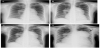
A 59-year-old woman was admitted to our hospital in a state of shock. She was diagnosed with SLE at 24 years of age, but had refused to take steroids or immunosuppressants. A diagnosis of pulmonary hypertension due to SLE was made at 42 years old. She continued to refuse certain kinds of medication, including steroids and immunosuppressants. The pulmonary artery increased in size over a decade (Figures 1A to 1C).
The patient had been relatively well until approximately 2 months before admission, when shortness of breath developed. Home oxygen therapy was introduced at 0.5 liters per minute of oxygen through a nasal cannula at rest or during sleep and 1 liter per minute of oxygen on effort, with some improvement. Approximately 1 month before admission, anasarca developed. Additional diuretics were administered, but little improvement was observed. Finally, she agreed to take prednisone 18 days before admission. Two days before admission, her appetite decreased and coldness of the limbs developed. On the day of admission, she had 2 brief episodes of weakness and was transferred to the emergency department of our hospital.
She reportedly had allergies to pyrazolone derivatives, beraprost sodium, bosentan hydrate, and sildenafil citrate. Her medical history was otherwise unremarkable. She did not drink alcohol, smoke, or use illicit drugs. Her medications included tadalafil at 20 mg (administered 2 months previously), digoxin at 0.25 mg, bisoprolol at 2.5 mg, tolvaptan at 15 mg, azosemide at 60 mg, and prednisone at 15 mg.
On admission, she was drowsy and appeared uncomfortable. Her blood pressure was 61/18 mm Hg, pulse was 126 beats per minute and irregular, body temperature was 34.4°C, respiratory rate was 18 breaths per minute, and oxygen saturation was 94% while breathing 4 liters of oxygen per minute through a face mask. The jugular veins were distended; a loud second heart sound and a systolic ejection murmur were audible without gallop rhythm; the respiratory sounds were diminished bilaterally at the lower lung fields but almost clear; the abdomen was soft without organomegaly or masses; and severe leg edema was present on both sides.
An electrocardiogram showed atrial fibrillation with a rate of 132 beats per minute, right axis deviation, right bundle branch block, and inverted T waves in the inferior leads. A chest radiograph showed mild pulmonary congestion with a cardiothoracic ratio of 81% and enlargement of the pulmonary artery with prominent protrusion of the second left arch (Figure 1D). The blood levels of total bilirubin and direct bilirubin were 6.7 mg/dl and 4.1 mg/dl. Creatinine and blood urea nitrogen levels were also elevated to 2.41 mg/dl and 84 mg/dl. The level of N-terminal pro-brain natriuretic peptide was 13,168 pg/ ml (reference range, ≤55).
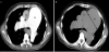
Transthoracic echocardiography was performed. The parasternal short-axis view of the echocardiogram revealed flattening of the ventricular septum, a dilated pulmonary artery and branches, and an enlarged right atrium and right ventricle. The systolic pulmonary artery pressure was estimated to be 60 mm Hg, similar to the systolic blood pressure. Computed tomography of the chest without contrast showed that the main pulmonary artery had increased in size compared with previous findings (Figure 2A), up to 82 mm in diameter (Figure 2B).
This patient died hours after admission, with no response to cardiopulmonary resuscitation. Autopsy showed an enlarged right ventricle and pulmonary alveolar hemorrhage with the formation of a plexiform lesion, accompanied by thrombi, in the branches of the pulmonary artery (Figure 3).

3. Discussion
The present patient died approximately 35 years after the diagnosis of SLE and 17 years after the diagnosis of pulmonary hypertension due to SLE. She had refused to take steroids or immunosuppressants for almost the entire disease duration. Furthermore, her allergies to most prostanoids, endothelin receptor antagonists, and phosphodiesterase- 5-inhibitors had made the treatment of pulmonary hypertension more difficult. We may safely consider changes of the pulmonary artery in this case as the natural course of pulmonary hypertension due to SLE.
Systematic reviews of patients with SLE [1-4] indicate that the incidence of pulmonary hypertension is 0.5% to 23.3% and that the diagnosis of pulmonary hypertension occurs 4.9 years to 10.7 years after the initial diagnosis of SLE. The diagnosis of pulmonary hypertension in our case was made 18 years after the diagnosis of SLE. Patients with pulmonary hypertension due to SLE seem to have a poorer outcome than patients with idiopathic pulmonary hypertension [5]. The mean survival period is reported to be from 13 months to 5 years in patients with pulmonary hypertension and SLE [1,5,6]. These findings suggest that our patient survived for a relatively long time (i.e., 17 years) despite a lack of appropriate medication, although the use of immunosuppressive therapy is controversial in patients with pulmonary hypertension and SLE [1,3].
The underlying mechanism linking SLE with pulmonary hypertension has not been fully elucidated, but proposed conditions include a hypercoagulable state (e.g., thrombotic arteriopathy), pulmonary vasculopathy (e.g., plexiform lesions), and immunemediated vasculopathy (e.g., pulmonary vasculitis) [1]. All of these 3 factors were present in our patient. Furthermore, acute diffuse alveolar damage and hemorrhage were confirmed on autopsy, features consistent with a common cause of death in patients with pulmonary hypertension due to SLE [7]. The maximum diameter of the main pulmonary artery was 82 mm in our patient. Complications leading to sudden death in patients with pulmonary artery aneurysm include pulmonary artery rupture, pulmonary artery dissection, and left main compression syndrome [8], which were not observed in our patient.
Competing Interests
The authors have declared that no competing interests exist.
Author Contributions
Yosuke Maehara and Tatsuya Kawasaki wrote the article and performed a literature review. Shigeyuki Miki, Yasuyuki Enoki, and Tadaaki Kamitani assisted with revisions. All authors read and approved this final manuscript.