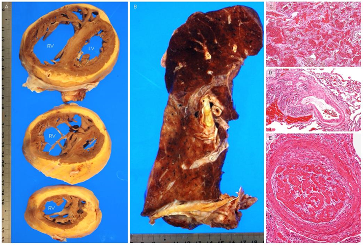
Figure 3: Autopsy findings.
The right ventricle is enlarged at basal, middle, and apical levels (A). Diffuse hemorrhage is suspected, especially in the upper field of the left lung (B). Pathological evaluation shows diffuse alveolar hemorrhage (C), plexiform lesions (D), and thrombi (E) in small and medium-sized arteries (hematoxylin and eosin). LV = left ventricle; RV = right ventricle.
The right ventricle is enlarged at basal, middle, and apical levels (A). Diffuse hemorrhage is suspected, especially in the upper field of the left lung (B). Pathological evaluation shows diffuse alveolar hemorrhage (C), plexiform lesions (D), and thrombi (E) in small and medium-sized arteries (hematoxylin and eosin). LV = left ventricle; RV = right ventricle.