


1. Introduction
Cephalohematoma in the neonate results from accumulation of blood between the bone and periosteum. The incidence is reported as 0.4% to 2.5 % in previous literature [1]. Bacterial infection is rarely complicated with a cephalohematoma and it is usually associated with sepsis, meningitis, or osteomyelitis of the underlying skull bone. Escherichia coli is the most common organism to be encountered, whereas Staphylococcus aureus, coagulase-negative staphylococci, Streptococcus pneumoniae, Bacillus spp., Pseudomonas spp., Proteus spp., Salmonella spp., Gardnerella vaginalis, group B streptococcus, and anaerobes are less common pathogens [2-6]. A recent study further highlights the polymicrobial nature and potential importance of anaerobic bacteria in infected cephalohematoma (IC) in neonates [7]. Gram negative rods represent the predominant bacteria of IC with concurrent meningitis in several literature reviews [1,2,8,12,17]. With antibiotic therapy and surgical drainage, we describe a neonate with huge IC with concomitant meningitis, recovered without any sequela.
2. Case report
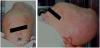
A 13-day-old, full-term, male neonate who was born via spontaneous vaginal delivery without vacuum extraction suffered from progressive enlargement of his occipital cephalohematoma for one week. He became febrile and irritable since 2 days prior to admission. On examination, a huge cephalohematoma (18cm in the greatest dimension) with fluctuation and local heat was noted (figure 1). The overlying skin was erythematous but intact. His anterior fontanel was neither tense nor bulged. The other physical and neurological examinations were unremarkable. A complete blood count revealed white blood cell (WBC) count of 13,400/mm3 with left shift. The C-reactive protein (CRP) level was 9.86 mg/dL. Intravenous ampicillin (150 mg/kg/day) and gentamicin (5 mg/ kg/day) were used as empiric antibiotic therapy after initial septic workup. As fever and irritability persisted on the second admission day, brain sonography was done with no intracranial lesion noted. The cerebrospinal fluid (CSF) study revealed WBC count of 340/ mm3 (with 8% neutrophils, 82% lymphocytes, and 8% monocytes), red blood cell (RBC) count of 50/ mm3, glucose level of 32 mg/ dL, and total protein level of 137 mg/dL. With the consideration of concomitant meningitis, intravenous ceftriaxone (100 mg/kg/day) therapy was started. The bacterial culture of blood and CSF yielded no microorganism. Low grade fever persisted and the cephalohematoma enlarged progressively. He received incision and drainage and became afebrile on the next day. The evacuated pus culture grew E. coli, which is susceptible to amipicillin, gentamicin, and ceftriaxone. Totally, he received intravenous ceftriaxone therapy for 21 days and no sequela is reported.
3. Discussion
An infected cephalohematoma is rare but potentially lifethreatening condition in neonates. Meningitis is rarely reported to be associated with IC in previous literature. In Taiwan, Chang et al. reported 3 of 28 infants with IC to be associated with meningitis in a period of 25 years [3]. A recent literature review reported 11 of 43 infants had IC with concurrent meningitis. The causative bacteria are predominately gram negative rods, especially E. coli [8]. The overall mortality rate remains high and which is related to the delayed diagnosis of meningitis and lack of surgical drainage [3,5,8-12].
The association of meningitis might result from bacteremic seeding or direct extension through the infected skull bone [13,14]. Focal extension accounts for the association of underlying skull bone osteomyelitis with negative blood culture and concurrent meningitis. Radiographic studies and bone scan may provide useful information in the differentiation of underlying skull bone osteomyelitis [8,13,14].
In the current case, delayed CSF study with previous exposure to antibiotics leads to the sterile result. Early detection of the association of meningitis is crucial, because prolonged therapy of antimicrobial agents with good tissue penetration is necessary for successful treatment. If such association is ignored, serious complication, including mortality, may ensue [15-17]. It is suggested CSF study is indicated for neonates with IC [5].
The current case received surgical drainage and antibiotic therapy for a total of 3 weeks. The adequate durations of antibiotic therapy have not yet been established. However, it is suggested that neonates with IC and concurrent bacteremia, meningitis, and osteomyelitis, to receive antibiotic therapy for at least 2-3 weeks, 3-4 weeks, and 4-6 weeks, respectively [6,15]. Surgical drainage plays an important role in the management of IC and failure of treatment has been reported with antimicrobial therapy alone [4,8-10, 16-18]. Repeated needle aspiration is also found to be a safe and effective alternative procedure for IC in neonates [6].
4. Conclusion
In conclusion, meningitis should be considered in neonate with IC. It necessitates appropriate selection of antibiotics with ability to penetrate blood-brain barrier and a prolonged period of therapy. Early detection of such association is crucial and related to their prognosis. Therefore, we suggest that CSF study is warranted for initial workup in such patients, if no contraindication exists. Further studies are necessary to confirm our findings and suggestions.
Competing Interests
The author declare that there is no competing interests regarding the publication of this article.