


1. Introduction
In medical practice, physicians in the field of internal medicine generally apply diagnostic imaging such as chest radiography, abdominal ultrasonography as their initial image inspection because it can easily obtain detailed internal information from a patient’s body. Interpreting images accurately and detecting diseases as early as possible is a decisive factor for physicians in providing effective subsequent treatment. However, professional image interpretation ability differs widely among physicians, and patients are at risk of disease aggravation due to medical malpractice caused by physicians’ failure in noticing abnormal findings. We analyzed a huge amount of knowledge, experiential information and actual case data stored at university hospitals and succeeded in developing data models ideal for teaching an AI (artificial intelligence) computer the knowledge, experience and case data required for image diagnosis by applying knowledge information processing technology [1]. Our "Diagnostic Imaging Knowledge Service" developed by using the above data models is an online service in which an AI computer provides the knowledge, experiential information and case images essential for image diagnosis with the aim of allowing general healthcare professionals to efficiently master the same image diagnosis process that specialists in image diagnosis generally apply to image interpretation in their daily medical practice. The online service has proven useful for a wide range of purposes such as clinical training of physicians and education of radiological technologists. The online service is also applicable as a tool to assist physicians in swiftly making the right decision when they encounter challenging cases during their daily medical practice.
2. Methods
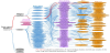
We analyzed an enormous number of cases stored at university hospitals to teach an AI computer the knowledge and experience required for image diagnosis. These analytical results revealed that when specialists in image diagnosis discover an abnormality in inspection images, they estimate the possibility of every conceivable disease occurring based on their past clinical experience and a combination of the following information: the location (body region), the findings (basic finding), and the supplemental findings (additional finding) of the abnormality. We then extracted the elements required for image diagnosis such as “body region”, “basic finding”, “additional finding”, and “diagnosis” as words, and numerically associated the combination of elements and the strength of their mutual combination with each other so that these numerical data accurately reflect the data obtained from actual past cases, the information obtained from literature published in the past, and medical specialists’ experience. We also associated prefixes and suffixes related to each element (word) with the extracted word as detailed elements. By way of these tasks, we succeeded in structuring and defining the knowledge, and experiential information required for image diagnosis, into a data format that an AI computer program can process. Figure 1 shows a schematic chart of the "diagnostic imaging knowledge base" for chest X-ray images defined with the above-mentioned tasks.
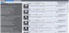
On the other hand, among a huge number of cases stored at university hospitals, we extracted cases likely to render a high learning effect. By using these cases, we prepared a "case database" consisting of case images, medical information on the cases which is the gender, age and definitive diagnosis, and medical records structured based on the above-mentioned "diagnostic imaging knowledge base" (Figure 2). Installation of the "diagnostic imaging knowledge base" and "case data base" in an AI computer program allowed it to effectively provide the knowledge, experiential information and case images required for evidence-based image diagnosis.
We created an online service called the “Diagnostic Imaging Knowledge Service" that incorporates this "diagnostic imaging knowledge base" and "case database" and that provides the following functions:
- Navigating users to select diagnosis based on data evidence from university hospitals;
- Allowing users to search case images leading to an accurate “diagnosis” and providing users with explanations given by specialists;
- Supporting users in preparing medical records through a simple procedure;
- Providing disease explanations which are concise and easy to understand even for patients;
- Providing an effective learning simulator that allows users to get a pseudo-experience of the same image diagnostic process that specialists generally apply to image interpretation in their own medical practice.
We would be grateful if you could take the time to check the website of the non-profit organization Medical Shinansha for details: https://www.medicalshinansha.or.jp/en/.
3. Results & Discussion
Healthcare professionals have conventionally learned how to interpret diagnostic images by receiving one-to-one instruction from supervisory doctors or referring to past published literature or in other words by ordinary learning methods. However, conventional learning methods have several drawbacks including for example that few supervisory doctors are available and teaching materials for diagnostic-image learning are inadequate. These drawbacks have caused the problem that professional ability in image interpretation differs widely among physicians. The problem still remains unsolved. The “Diagnostic Imaging Knowledge Service" however, proves superior to the conventional learning methods in the following points:
- Ensuring the quality and quantity of the instruction content, because the “Diagnostic Imaging Knowledge Service" was developed based on knowledge, experiential information, and actual case data stored at university hospitals;
- Ensuring the quality and comprehensiveness of case images;
- Being available online.
As a result, the “Diagnostic Imaging Knowledge Service" has been in use within Japan and overseas as follows:
- Hyogo Prefectural Kakogawa Medical Center has made use of the “Diagnostic Imaging Knowledge Service" in clinical training since April 2018;
- Suzuka University of Medical Science has made use of the “Diagnostic Imaging Knowledge Service" in training of radiological technologists since October 2019;
- The Philippine College of Chest Physicians has made use of the "Diagnostic Imaging Tutor" which is an English version of the “Diagnostic Imaging Knowledge Service" in clinical training since December 2020.
The “Diagnostic Imaging Knowledge Service" has also proved effective in training of radiological technologists [2].
Receiving full recognition for its excellent performance from both academic and social viewpoints has led to the “Diagnostic Imaging Knowledge Service" receiving awards and being selected as the subject for promotion projects and commissioned projects as follows:
- Granted the Civil Society Organization (CSO) Award 2017 of Osaka Mayor Award in October 2017;
- Granted the Outstanding Presentation Award in the 37th Joint Conference on Medical Informatics in June 2018;
- Selected as the subject for the Welfare and Medical Promotion Project and the Osaka City Citizenship Promotion Project in fiscal 2019;
- Selected as the subject for the Program for capacity building on the quality of medical imaging and its diagnosis in the Philippines in fiscal 2019 (ongoing program).
4. Conclusions
In medical practice, diagnostic imaging is an effective tool allowing early detection and effective treatment of diseases because healthcare professionals can easily obtain detailed internal information from a patient’s body. However, professional ability in image interpretation differs widely among healthcare professionals. We analyzed an enormous number of cases stored at university hospitals. By way of analytical data, we developed the "Diagnostic Imaging Knowledge Service" by structuring and defining the knowledge, experiential information and actual case data required for image diagnosis, into a data format that an AI computer program can process. We have provided the "Diagnostic Imaging Knowledge Service" as an online service with the aim of allowing healthcare professionals to efficiently learn and master image diagnosis.
The "Diagnostic Imaging Knowledge Service" has been effectively utilized both inside and outside Japan because the "Diagnostic Imaging Knowledge Service" proves superior to conventional learning methods. A high rating of its excellent performance from both academic and social viewpoints resulted in the "Diagnostic Imaging Knowledge Service" being selected as the subject for promotion and commissioned projects within Japan and overseas.
We expect that the "Diagnostic Imaging Knowledge Service" will enhance image diagnosis to a yet higher level, facilitate early detection of diseases, and contribute to reducing the physical, economic and mental burdens imposed not only on healthcare professionals but also on patients and their families.
Competing Interests
The author declare that there is no competing interests regarding the publication of this article.