


1. Introduction
Pharyngeal inverted papilloma is a rare lesion. Inverted papillomas commonly originate in the sinonasal cavity, with human papilloma virus (HPV) infection being a possible causative factor. Sinonasal inverted papilloma is generally benign and has widely been investigated for its features, etiology, and treatment. On the other hand, extranasal inverted papilloma is rare and can involve the pharynx [1-8], ear [9], and cervical lymph nodes [10]. Inverted papilloma originates from the Schneiderian mucosa, which covers the nasal cavity [11], possibly explaining why this lesion frequently involves the sinonasal cavity and rarely involves the extranasal regions. Ectopic migration into extranasal areas of the Schneiderian mucosa is believed to be attributed to the development of extranasal inverted papilloma [11]. Papilloma of the pharynx is commonly known as exophytic papilloma; however, pharyngeal inverted papilloma is rarely reported [1-8], with no literature reviews. Consequently, the features of this lesion, including the sites of involvement, malignant transformation rate, and recurrence rate, remain to be elucidated. To the best of our knowledge, we present the first case of an extranasal pharyngeal inverted papilloma with parapharyngeal space invasion and associated hearing loss and the first literature review on the features of this lesion.
2. Case Presentation
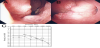
A 77-year-old man presented with left-sided hearing loss and sore throat of 1 month’s duration. On examination, there was no tinnitus, vertigo, fever, cough, or neurological deficit. His left tympanum appeared dull, suggesting otitis media with effusion. Fiberscopy of the ears, nose, and throat (ENT) revealed an irregular mass over the inferior concha, nasal floor, and nasal septum in the right nasal cavity (Figure 1a) and in the fossa of Rosenmüller and the anterior nasopharynx adjacent to the Eustachian canal in the left nasopharynx (Figure 1b). Pure-tone audiography revealed mixed conductive– sensorineural hearing loss on the left side. The average air- and boneconduction hearing levels at 500, 1000, and 2000 Hz were 61.7 dB and 53.3 dB, respectively (Figure 1c). Soft palate elevation was satisfactory, and both vocal cords were adequately mobile. Other cranial nerve functions were intact.
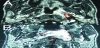
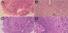
Enhanced T1-weighted magnetic resonance imaging showed a high-intensity area in the left nasopharynx, including the fossa of Rosenmüller and parapharyngeal space (Figure 2), and the right inferior concha. Pathological examination of a biopsy specimen from the right nasal cavity revealed endophytic, multilayered, proliferating squamous epithelium (Figure 3a) and koilocytosis, suggesting viral infection (Figure 3b). A specimen from the left nasopharynx also showed endophytic, multilayered, proliferating squamous epithelium (Figure 3c) and prominent koilocytosis (Figure 3d). On the basis of these findings, the masses in the right nasal cavity (Figure 3a,b) and left nasopharynx (Figure 3c,d) were diagnosed as right nasal inverted papilloma and left nasopharyngeal inverted papilloma, respectively. Surgical resection was suggested; however, the patient requested conservative management without surgery. After clarithromycin 400 mg and carbocisteine 750mg were administered for 2 weeks, otitis media with effusion was cured. During 3-month follow-up periods, tumor did not proliferate.
3. Discussion
The male: female ratio for the incidence of pharyngeal inverted papilloma is 9:4 (Table 1). The average age of patients has been 58.3 years (range, 31–79 years). Pharyngeal inverted papilloma occurs most commonly in the nasopharynx (65.4%), followed by the oropharynx (30.8%) and hypopharynx (3.8%)(Table 1). The symptoms include sore throat (21.7%; 5 of 23), foreign body sensation (17.4%; 4 of 23), hearing loss (17.4%; 4 of 23), dysphagia (8.7%; 2 of 23), otalgia (4.3%; 1 of 23), nasal obstruction (4.3%; 1 of 23), nasal speech (4.3%; 1 of 23), and rhinorrhea (4.3%; 1 of 23); 52.2% (12 of 23) patients were asymptomatic. Hearing loss occurred only in association with nasopharyngeal inverted papilloma, probably because the function of the Eustachian canal is easily disturbed by a nasopharyngeal tumor.
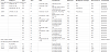
Surprisingly, approximately 50% patients are diagnosed during other examinations or treatments, such as endoscopy or sinus surgery. The difference in incidence among sites can be attributed to anatomical specifications. Sinonasal inverted papilloma originates from the Schneiderian membrane covering the nasal cavity except the nasal vestibule and the superior wall of the nasal cavity [11]. The Schneiderian membrane is a nasal mucosal layer of ciliated columnar epithelium with an ectodermal origin [11]. Ectopic migration of this membrane to the pharynx occurs in the order of the nasopharynx, oropharynx, and hypopharynx, considering the distance of each segment from the nasal cavity.
These lesions are generally excised by surgery; however, our review found a recurrence rate of 31.6% (6 of 19), which is considerably higher than that for sinonasal inverted papilloma (endoscopically treated cases, 12%; nonendoscopically treated cases, 20%) [12]. These differences in recurrence rates may be caused by differences in surgical approaches. For example, the pharynx, particularly the nasopharynx, is surrounded by vital structures; therefore, radical excision is impossible. Another reason may be differences in the causative virus.
Although pharyngeal inverted papilloma is benign, it can show potential malignant transformation, similar to sinonasal inverted papilloma. Our review revealed a malignant transformation rate of 15.4% (4 of 26). The pathological diagnosis was squamous cell carcinoma in three cases and Schneiderian carcinoma in one. The reported rate of malignant transformation for sinonasal inverted papilloma is considerably lower at 8% [13]. Sinonasal inverted papilloma is commonly related to HPV 6, 11, 16, and 18 [14], with the risk of malignant transformation being low with the former two and high with the latter two [14]. HPV is also believed to play a role in the etiology of pharyngeal inverted papilloma. Koilocytes, which are cells with vacuolization around the nucleus and suggest HPV infection, were observed in pathology specimens from our patient and a previous patient [3]. However, to date, HPV DNA has not been investigated in pharyngeal inverted papilloma.
HPV induces pharyngeal carcinoma and is most frequently detected in patients with oropharyngeal carcinoma [15], with HPV 16 being the most common. On the other hand, nasopharyngeal and hypopharyngeal carcinomas are rarely associated with HPV [15]. The most common sites of involvement differ between HPV-related pharyngeal carcinoma and pharyngeal inverted papilloma, possibly because of differences in the virus type. Therefore, the types of HPV should be further investigated for clarification of our findings.
4. Conclusion
In conclusion, we present the case of a patient with nasopharyngeal inverted papilloma accompanied by otitis media with effusion, loss, and sore throat. The tumor invaded the parapharyngeal space and disturbed the function of the Eustachian canal. Histopathology confirmed an inverted papilloma with koilocytes, suggesting HPV infection. According to our literature review, the most common site of involvement is the nasopharynx, followed by the oropharynx and hypopharynx. Koilocytes were observed in our patient and a previously reported patient, suggesting the contribution of HPV infection. However, HPV DNA in such cases has not been investigated; further studies are necessary to elucidate the etiology.
Competing Interests
The author declare that he has no competing interests.