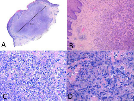
Figure 2: A (sub-macroscopic view) – B (1X): irregularly shaped nodule with central ulceration and diffuse infiltration of large tumor cells into the dermis. C (20X) – D (40X): tumor cells exhibited an irregular shape with abundant eosinophilic, sometimes clear, cytoplasm and large, irregular shaped nuclei with a lobulated or dented appearance.