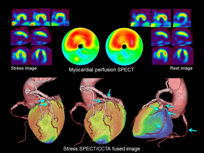
Figure 6: Case of 70-year-old man with effort angina pectoris who has history of myocardial infarction and CABG (LITA-LAD, Ao- SVG-PL, and GEA-#4PD). Coronary angiography showed a CTO in the mid RCA, a 99% stenosis in the proximal-mid LAD, a 90% stenosis in the proximal LCX, a CTO in the mid LCX, and a 90% stenosis in the PL. All the bypass grafts were patent. Exercise-stress myocardial perfusion SPECT using 99mTc-tetrofosmin (non-AC images) showed myocardial ischemia only in the anterior wall. On hybrid SPECT/CT, the anterior myocardial ischemia was located in both the first and second diagonal branch territories and the stenotic lesion in the proximal LAD was confirmed to be a culprit lesion.