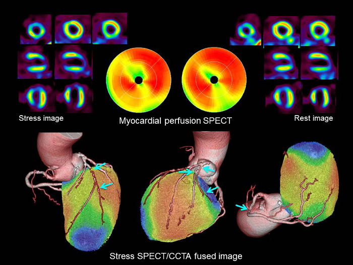
Figure 5: Case of 78-year-old woman with multivessel disease and chronic total occlusion. CCTA revealed a >75% stenosis in the mid RCA, a chronic total occlusion in the proximal–mid LAD, a >90% stenosis in the first diagonal branch, a >75% stenosis in the proximal LCX, a >75% stenosis in the obtuse marginal branch, and a >75% stenosis in the mid LCX. Adenosine-stress myocardial perfusion SPECT using 99mTc-tetrofosmin (non-AC images) showed a partially reversible perfusion defect in the anteroapical wall and a fixed defect in the inferoposterior wall. On hybrid SPECT/CT, the anteroapical myocardial ischemia was located in the LAD and first diagonal branch territory and the inferoposterior myocardial infarction was situated in the RCA territory.