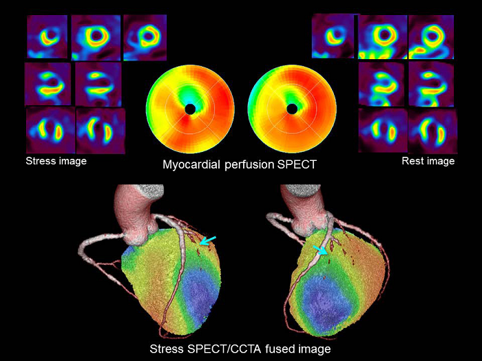
Figure 4: Case of 73-year-old man with branch disease and silent myocardial ischemia who has history of PCI in RCA and LAD. CCTA revealed a >90% stenosis in the first diagonal branch. Exercise-stress myocardial perfusion SPECT using 99mTc-tetrofosmin (non-AC images) showed a partially reversible perfusion defect in the anteroapical wall. On hybrid SPECT/CT, the anteroapical ischemia was located in the first diagonal branch territory, while the LAD territory did not show any perfusion defects.